[English] 日本語
Yorodumi Papers
- Database of articles cited by EMDB/PDB/SASBDB data -

+
Search query

Keywords
Structure methods
Author
Journal
IF

-
Structure paper

TitleStructures of the mitochondrial single-stranded DNA binding protein with DNA and DNA polymerase γ.
Journal, issue, pagesNucleic Acids Res, Vol. 52, Issue 17, Page 10329-10340, Year 2024
Publish dateSep 23, 2024
AuthorsAmanda A Riccio / Jonathan Bouvette / Lars C Pedersen / Shruti Somai / Robert C Dutcher / Mario J Borgnia / William C Copeland /
PubMed AbstractThe mitochondrial single-stranded DNA (ssDNA) binding protein, mtSSB or SSBP1, binds to ssDNA to prevent secondary structures of DNA that could impede downstream replication or repair processes. ...The mitochondrial single-stranded DNA (ssDNA) binding protein, mtSSB or SSBP1, binds to ssDNA to prevent secondary structures of DNA that could impede downstream replication or repair processes. Clinical mutations in the SSBP1 gene have been linked to a range of mitochondrial disorders affecting nearly all organs and systems. Yet, the molecular determinants governing the interaction between mtSSB and ssDNA have remained elusive. Similarly, the structural interaction between mtSSB and other replisome components, such as the mitochondrial DNA polymerase, Polγ, has been minimally explored. Here, we determined a 1.9-Å X-ray crystallography structure of the human mtSSB bound to ssDNA. This structure uncovered two distinct DNA binding sites, a low-affinity site and a high-affinity site, confirmed through site-directed mutagenesis. The high-affinity binding site encompasses a clinically relevant residue, R38, and a highly conserved DNA base stacking residue, W84. Employing cryo-electron microscopy, we confirmed the tetrameric assembly in solution and capture its interaction with Polγ. Finally, we derived a model depicting modes of ssDNA wrapping around mtSSB and a region within Polγ that mtSSB binds.
External linksNucleic Acids Res / PubMed:39106165 / PubMed Central
MethodsEM (single particle) / X-ray diffraction
Resolution1.9 - 3.5 Å
Structure data

EMDB-42842: A Mitochondrial Replication Complex: PolG/PolG2 Bound to DNA in complex with the Single-Stranded Binding Protein (mtSSB)
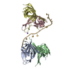
Method: EM (single particle) / Resolution: 3.5 Å

PDB-8uzt:
Mitochondrial single-stranded binding protein bound to DNA
Method: X-RAY DIFFRACTION / Resolution: 1.9 Å

Chemicals

ChemComp-GOL:
GLYCEROL

ChemComp-NA:
Unknown entry

ChemComp-HOH:
WATER

Source
  • homo sapiens (human)
  • synthetic construct (others)
KeywordsDNA BINDING PROTEIN/DNA / single-stranded binding protein / DNA BINDING PROTEIN / mitochondrial SSB / SSB / mitochondrial replication protein / DNA BINDING PROTEIN-DNA complex

+
About Yorodumi Papers

-
News

-
Feb 9, 2022. New format data for meta-information of EMDB entries

New format data for meta-information of EMDB entries

  • Version 3 of the EMDB header file is now the official format.
  • The previous official version 1.9 will be removed from the archive.

Related info.:EMDB header

External links:wwPDB to switch to version 3 of the EMDB data model

-
Aug 12, 2020. Covid-19 info

Covid-19 info

URL: https://pdbj.org/emnavi/covid19.php

New page: Covid-19 featured information page in EM Navigator.

Related info.:Covid-19 info / Mar 5, 2020. Novel coronavirus structure data

+
Mar 5, 2020. Novel coronavirus structure data

Novel coronavirus structure data

Related info.:Yorodumi Speices / Aug 12, 2020. Covid-19 info

External links:COVID-19 featured content - PDBj / Molecule of the Month (242):Coronavirus Proteases

+
Jan 31, 2019. EMDB accession codes are about to change! (news from PDBe EMDB page)

EMDB accession codes are about to change! (news from PDBe EMDB page)

  • The allocation of 4 digits for EMDB accession codes will soon come to an end. Whilst these codes will remain in use, new EMDB accession codes will include an additional digit and will expand incrementally as the available range of codes is exhausted. The current 4-digit format prefixed with “EMD-” (i.e. EMD-XXXX) will advance to a 5-digit format (i.e. EMD-XXXXX), and so on. It is currently estimated that the 4-digit codes will be depleted around Spring 2019, at which point the 5-digit format will come into force.
  • The EM Navigator/Yorodumi systems omit the EMD- prefix.

Related info.:Q: What is EMD? / ID/Accession-code notation in Yorodumi/EM Navigator

External links:EMDB Accession Codes are Changing Soon! / Contact to PDBj

+
Jul 12, 2017. Major update of PDB

Major update of PDB

  • wwPDB released updated PDB data conforming to the new PDBx/mmCIF dictionary.
  • This is a major update changing the version number from 4 to 5, and with Remediation, in which all the entries are updated.
  • In this update, many items about electron microscopy experimental information are reorganized (e.g. em_software).
  • Now, EM Navigator and Yorodumi are based on the updated data.

External links:wwPDB Remediation / Enriched Model Files Conforming to OneDep Data Standards Now Available in the PDB FTP Archive

-
Yorodumi Papers

Database of articles cited by EMDB/PDB/SASBDB data

  • Database of articles cited by EMDB, PDB, and SASBDB entries
  • Using PubMed data

Related info.:EMDB / PDB / SASBDB / Yorodumi / EMN Papers / Changes in new EM Navigator and Yorodumi

Read more