Movie
Movie Controller
 Structure viewers
 About Yorodumi Papers


+Search query
-Structure paper
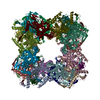
| Title | Molecular architecture of the mammalian 2-oxoglutarate dehydrogenase complex. |
|---|---|
| Journal, issue, pages | Nat Commun, Vol. 15, Issue 1, Page 8407, Year 2024 |
| Publish date | Sep 27, 2024 |
 Authors Authors | Yitang Zhang / Maofei Chen / Xudong Chen / Minghui Zhang / Jian Yin / Zi Yang / Xin Gao / Sensen Zhang / Maojun Yang /   |
| PubMed Abstract | The 2-oxoglutarate dehydrogenase complex (OGDHc) orchestrates a critical reaction regulating the TCA cycle. Although the structure of each OGDHc subunit has been solved, the architecture of the ...The 2-oxoglutarate dehydrogenase complex (OGDHc) orchestrates a critical reaction regulating the TCA cycle. Although the structure of each OGDHc subunit has been solved, the architecture of the intact complex and inter-subunit interactions still remain unknown. Here we report the assembly of native, intact OGDHc from Sus scrofa heart tissue using cryo-electron microscopy (cryo-EM), cryo-electron tomography (cryo-ET), and subtomogram averaging (STA) to discern native structures of the whole complex and each subunit. Our cryo-EM analyses revealed the E2o cubic core structure comprising eight homotrimers at 3.3-Å resolution. More importantly, the numbers, positions and orientations of each OGDHc subunit were determined by cryo-ET and the STA structures of the core were resolved at 7.9-Å with the peripheral subunits reaching nanometer resolution. Although the distribution of the peripheral subunits E1o and E3 vary among complexes, they demonstrate a certain regularity within the position and orientation. Moreover, we analyzed and validated the interactions between each subunit, and determined the flexible binding mode for E1o, E2o and E3, resulting in a proposed model of Sus scrofa OGDHc. Together, our results reveal distinctive factors driving the architecture of the intact, native OGDHc. |
 External links External links | Nat Commun / PubMed:39333186 / PubMed Central |
| Methods | EM (single particle) / EM (subtomogram averaging) / EM (tomography) |
| Resolution | 3.3 - 12.2 Å |
| Structure data | EMDB-37965, PDB-8x02:  EMDB-37966: E1 of 2-oxoglutarate dehydrogenase complex subtomogram averaging map  EMDB-37967: E2 of 2-oxoglutarate dehydrogenase complex subtomogram averaging map  EMDB-37968: E3 of 2-oxoglutarate dehydrogenase complex subtomogram averaging map  EMDB-61087: in situ E1 of 2-oxoglutarate dehydrogenase complex subtomogram averaging map  EMDB-61089: in situ E2 of 2-oxoglutarate dehydrogenase complex subtomogram averaging map  EMDB-61090: in situ E3 of 2-oxoglutarate dehydrogenase complex subtomogram averaging map |
| Source |
|
 Keywords Keywords | TRANSFERASE / Structure of mitochondrial enzyme complex |