 Movie
Movie Controller
 Structure viewers
 About Yorodumi Papers


+Search query
-Structure paper
| Title | Native structure of a retroviral envelope protein and its conformational change upon interaction with the target cell. |
|---|---|
| Journal, issue, pages | J Struct Biol, Vol. 197, Issue 2, Page 172-180, Year 2017 |
| Publish date | Jun 23, 2016 |
 Authors Authors | Christiane Riedel / Daven Vasishtan / C Alistair Siebert / Cathy Whittle / Maik J Lehmann / Walther Mothes / Kay Grünewald /     |
| PubMed Abstract | Enveloped viruses enter their host cells by membrane fusion. The process of attachment and fusion in retroviruses is mediated by a single viral envelope glycoprotein (Env). Conformational changes of ...Enveloped viruses enter their host cells by membrane fusion. The process of attachment and fusion in retroviruses is mediated by a single viral envelope glycoprotein (Env). Conformational changes of Env in the course of fusion are a focus of intense studies. Here we provide further insight into the changes occurring in retroviral Env during its initial interaction with the cell, employing murine leukemia virus (MLV) as model system. We first determined the structure of both natively membrane anchored MLV Env and MLV Env tagged with YFP in the proline rich region (PRR) by electron cryo tomography (cET) and sub-volume averaging. At a resolution of ∼20Å, native MLV Env presents as a hollow trimer (height ∼85Å, diameter ∼120Å) composed of step-shaped protomers. The major difference to the YFP-tagged protein was in regions outside of the central trimer. Next, we focused on elucidating the changes in MLV Env upon interaction with a host cell. Virus interaction with the plasma membrane occurred over a large surface and Env clustering on the binding site was observed. Sub-volume averaging did yield a low-resolution structure of Env interacting with the cell, which had lost its threefold symmetry and was elongated by ∼35Å in comparison to the unbound protein. This indicates a major rearrangement of Env upon host cell binding. At the site of virus interaction, the otherwise clearly defined bilayer structure of the host cell plasma membrane was much less evident, indicative of integral membrane protein accumulation and/or a change in membrane lipid composition. |
 External links External links | J Struct Biol / PubMed:27345930 / PubMed Central |
| Methods | EM (subtomogram averaging) |
| Resolution | 15.0 - 22.0 Å |
| Structure data |  EMDB-3357:  EMDB-3363: 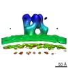 EMDB-3365: 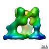 EMDB-3373: |
| Source |
|
 Murine leukemia virus