 Movie
Movie Controller
 Structure viewers
 About Yorodumi Papers


+Search query
-Structure paper
| Title | Hexadecameric structure of an invertebrate gap junction channel. |
|---|---|
| Journal, issue, pages | J Mol Biol, Vol. 428, Issue 6, Page 1227-1236, Year 2016 |
| Publish date | Mar 27, 2016 |
 Authors Authors | Atsunori Oshima / Tomohiro Matsuzawa / Kazuyoshi Murata / Kazutoshi Tani / Yoshinori Fujiyoshi /  |
| PubMed Abstract | Innexins are invertebrate-specific gap junction proteins with four transmembrane helices. These proteins oligomerize to constitute intercellular channels that allow for the passage of small signaling ...Innexins are invertebrate-specific gap junction proteins with four transmembrane helices. These proteins oligomerize to constitute intercellular channels that allow for the passage of small signaling molecules associated with neural and muscular electrical activity. In contrast to the large number of structural and functional studies of connexin gap junction channels, few structural studies of recombinant innexin channels are reported. Here we show the three-dimensional structure of two-dimensionally crystallized Caenorhabditis elegans innexin-6 (INX-6) gap junction channels. The N-terminal deleted INX-6 proteins are crystallized in lipid bilayers. The three-dimensional reconstruction determined by cryo-electron crystallography reveals that a single INX-6 gap junction channel comprises 16 subunits, a hexadecamer, in contrast to chordate connexin channels, which comprise 12 subunits. The channel pore diameters at the cytoplasmic entrance and extracellular gap region are larger than those of connexin26. Two bulb densities are observed in each hemichannel, one in the pore and the other at the cytoplasmic side of the hemichannel in the channel pore pathway. These findings imply a structural diversity of gap junction channels among multicellular organisms. |
 External links External links | J Mol Biol / PubMed:26883891 |
| Methods | EM (electron crystallography) |
| Resolution | 10.0 Å |
| Structure data |  EMDB-3345: 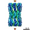 EMDB-3346: |
| Source |
|

