 Movie
Movie Controller
 Structure viewers
 About Yorodumi Papers


+Search query
-Structure paper
| Title | Three-Dimensional Reconstruction of the Hepatitis C Virus Envelope Glycoprotein E1E2 Heterodimer by Electron Microscopic Analysis. |
|---|---|
| Journal, issue, pages | J Virol, Vol. 97, Issue 1, Page e0178822, Year 2023 |
| Publish date | Jan 31, 2023 |
 Authors Authors | Tapan Kanai / Zongyi Hu / Renbin Yang / Weimin Wu / Ziqiu Wang / Christopher D Ma / Jaime Sanchez-Meza / Mansun Law / Michael Houghton / John Lokman Law / Michael Logan / Natalia de Val / T Jake Liang /   |
| PubMed Abstract | Despite the development of highly effective hepatitis C virus (HCV) treatments, an effective prophylactic vaccine is still lacking. HCV infection is mediated by its envelope glycoproteins, E1 and E2, ...Despite the development of highly effective hepatitis C virus (HCV) treatments, an effective prophylactic vaccine is still lacking. HCV infection is mediated by its envelope glycoproteins, E1 and E2, during the entry process, with E2 binding to cell receptors and E1 mediating endosomal fusion. The structure of E1E2 has only been partially resolved by X-ray crystallography of the core domain of E2 protein (E2c) and its complex with various neutralizing antibodies. Structural understanding of the E1E2 heterodimer in its native form can advance the design of candidates for HCV vaccine development. Here, we analyze the structure of the recombinant HCV E1E2 heterodimer with the aid of well-defined monoclonal anti-E1 and E2 antibodies, as well as a small-molecule chlorcyclizine-diazirine-biotin that can target and cross-link the putative E1 fusion domain. Three-dimensional (3D) models were generated after extensive 2D classification analysis with negative-stain single-particle data sets. We modeled the available crystal structures of the E2c and Fabs into 3D volumes of E1E2-Fab complexes based on the shape and dimension of the domain density. The E1E2 heterodimer exists in monomeric form and consists of a main globular body, presumably depicting the E1 and E2 stem/transmembrane domain, and a protruding structure representing the E2c region, based on anti-E2 Fab binding. At low resolution, a model generated from negative-stain analysis revealed the unique binding and orientation of individual or double Fabs onto the E1 and E2 components of the complex. Cryo-electron microscopy (cryo-EM) of the double Fab complexes resulted in a refined structural model of the E1E2 heterodimer, presented here. Recombinant HCV E1E2 heterodimer is being developed as a vaccine candidate. Using electron microscopy, we demonstrated unique features of E1E2 in complex with various neutralizing antibodies and small molecule inhibitors that are important to understanding its antigenicity and induction of immune response. |
 External links External links | J Virol / PubMed:36519897 / PubMed Central |
| Methods | EM (single particle) |
| Resolution | 6.8 - 30.0 Å |
| Structure data | 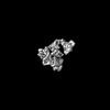 EMDB-29492: Three-Dimensional Reconstruction of HCV Envelope Glycoproteins E1E2 Heterodimer by Electron Microscopic Analysis  EMDB-29499: Three-Dimensional Reconstruction of HCV Envelope Glycoproteins E1E2 Heterodimer by Electron Microscopic Analysis |
| Source |
|
 Hepacivirus C