 Movie
Movie Controller
 Structure viewers
 About Yorodumi Papers


+Search query
-Structure paper
| Title | The structure of purified kinetochores reveals multiple microtubule-attachment sites. |
|---|---|
| Journal, issue, pages | Nat Struct Mol Biol, Vol. 19, Issue 9, Page 925-929, Year 2012 |
| Publish date | Aug 12, 2012 |
 Authors Authors | Shane Gonen / Bungo Akiyoshi / Matthew G Iadanza / Dan Shi / Nicole Duggan / Sue Biggins / Tamir Gonen /  |
| PubMed Abstract | Chromosomes must be accurately partitioned to daughter cells to prevent aneuploidy, a hallmark of many tumors and birth defects. Kinetochores are the macromolecular machines that segregate ...Chromosomes must be accurately partitioned to daughter cells to prevent aneuploidy, a hallmark of many tumors and birth defects. Kinetochores are the macromolecular machines that segregate chromosomes by maintaining load-bearing attachments to the dynamic tips of microtubules. Here, we present the structure of isolated budding-yeast kinetochore particles, as visualized by EM and electron tomography of negatively stained preparations. The kinetochore appears as an ~126-nm particle containing a large central hub surrounded by multiple outer globular domains. In the presence of microtubules, some particles also have a ring that encircles the microtubule. Our data, showing that kinetochores bind to microtubules via multivalent attachments, lay the foundation to uncover the key mechanical and regulatory mechanisms by which kinetochores control chromosome segregation and cell division. |
 External links External links | Nat Struct Mol Biol / PubMed:22885327 / PubMed Central |
| Methods | EM (tomography) |
| Structure data | 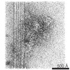 EMDB-2153: 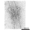 EMDB-2154: |
| Source |
|

