 Movie
Movie Controller
 Structure viewers
 About Yorodumi Papers


+Search query
-Structure paper
| Title | Molecular architecture of synaptic vesicles. |
|---|---|
| Journal, issue, pages | Proc Natl Acad Sci U S A, Vol. 121, Issue 49, Page e2407375121, Year 2024 |
| Publish date | Dec 3, 2024 |
 Authors Authors | Uljana Kravčenko / Max Ruwolt / Jana Kroll / Artsemi Yushkevich / Martina Zenkner / Julia Ruta / Rowaa Lotfy / Erich E Wanker / Christian Rosenmund / Fan Liu / Mikhail Kudryashev /  |
| PubMed Abstract | Synaptic vesicles (SVs) store and transport neurotransmitters to the presynaptic active zone for release by exocytosis. After release, SV proteins and excess membrane are recycled via endocytosis, ...Synaptic vesicles (SVs) store and transport neurotransmitters to the presynaptic active zone for release by exocytosis. After release, SV proteins and excess membrane are recycled via endocytosis, and new SVs can be formed in a clathrin-dependent manner. This process maintains complex molecular composition of SVs through multiple recycling rounds. Previous studies explored the molecular composition of SVs through proteomic analysis and fluorescent microscopy, proposing a model for an average SV (1). However, the structural heterogeneity and molecular architecture of individual SVs are not well described. Here, we used cryoelectron tomography to visualize molecular details of SVs isolated from mouse brains and inside cultured neurons. We describe several classes of small proteins on the SV surface and long proteinaceous densities inside SVs. We identified V-ATPases, determined a structure using subtomogram averaging, and showed them forming a complex with the membrane-embedded protein synaptophysin (Syp). Our bioluminescence assay revealed pairwise interactions between vesicle-associated membrane protein 2 and Syp and V-ATPase Voe1 domains. Interestingly, V-ATPases were randomly distributed on the surface of SVs irrespective of vesicle size. A subpopulation of isolated vesicles and vesicles inside neurons contained a partially assembled clathrin coat with an icosahedral symmetry. We observed V-ATPases under clathrin cages in several isolated clathrin-coated vesicles (CCVs). Additionally, from isolated SV preparations and within hippocampal neurons we identified clathrin baskets without vesicles. We determined their and CCVs preferential location in proximity to the cell membrane. Our analysis advances the understanding of individual SVs' diversity and their molecular architecture. |
 External links External links | Proc Natl Acad Sci U S A / PubMed:39602275 / PubMed Central |
| Methods | EM (subtomogram averaging) |
| Resolution | 15.6 - 172.5 Å |
| Structure data | 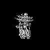 EMDB-18556: Structure of V-ATPase obtained from isolated mouse synaptic vesicles 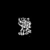 EMDB-18557: A membrane-embedded intravesicular stick-shaped protein located in proximity to V-ATPase  EMDB-18568: Clathrin triskelion from the coat assembled on synaptic vesicle membranes isolated from mouse brain tissue 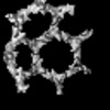 EMDB-18572: A segment of clathrin cage from the coat assembled on synaptic vesicle membranes isolated from mouse brain tissue 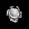 EMDB-18574: A segment of clathrin-coated endosome, isolated from mouse brain tissue 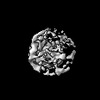 EMDB-18578: Non-vesicle carrying clathrin basket, isolated from mouse brain tissue 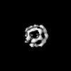 EMDB-18582: Non-vesicle carrying clathrin basket from primary hippocampal neurons, cultured on grids 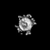 EMDB-18583: A segment of a clathrin-coated vesicle obtained from a primary hippocampal neurons, cultured on grids  EMDB-18584: Clathrin triskelion from the coat assembled on vesicle membranes in primary hippocampal neurons, cultured on grids |
| Source |
|

