 Movie
Movie Controller
 Structure viewers
 About Yorodumi Papers


+Search query
-Structure paper
| Title | In situ snapshots along a mammalian selective autophagy pathway. |
|---|---|
| Journal, issue, pages | Proc Natl Acad Sci U S A, Vol. 120, Issue 12, Page e2221712120, Year 2023 |
| Publish date | Mar 21, 2023 |
 Authors Authors | Meijing Li / Ishita Tripathi-Giesgen / Brenda A Schulman / Wolfgang Baumeister / Florian Wilfling /  |
| PubMed Abstract | Selective macroautophagy (hereafter referred to as autophagy) describes a process in which cytosolic material is engulfed in a double membrane organelle called an autophagosome. Autophagosomes are ...Selective macroautophagy (hereafter referred to as autophagy) describes a process in which cytosolic material is engulfed in a double membrane organelle called an autophagosome. Autophagosomes are carriers responsible for delivering their content to a lytic compartment for destruction. The cargo can be of diverse origin, ranging from macromolecular complexes to protein aggregates, organelles, and even invading pathogens. Each cargo is unique in composition and size, presenting different challenges to autophagosome biogenesis. Among the largest cargoes targeted by the autophagy machinery are intracellular bacteria, which can, in the case of range from 2 to 5 μm in length and 0.5 to 1.5 μm in width. How phagophores form and expand on such a large cargo remains mechanistically unclear. Here, we used HeLa cells infected with an auxotrophic to study the process of phagophore biogenesis using in situ correlative cryo-ET. We show that host cells generate multiple phagophores at the site of damaged -containing vacuoles (SCVs). The observed double membrane structures range from disk-shaped to expanded cup-shaped phagophores, which have a thin intermembrane lumen with a dilating rim region and expand using the SCV, the outer membrane of , or existing phagophores as templates. Phagophore rims establish different forms of contact with the endoplasmic reticulum (ER) via structurally distinct molecular entities for membrane formation and expansion. Early omegasomes correlated with the marker Double-FYVE domain-Containing Protein 1 (DFCP1) are observed in close association with the ER without apparent membrane continuity. Our study provides insights into the formation of phagophores around one of the largest selective cargoes. |
 External links External links | Proc Natl Acad Sci U S A / PubMed:36917659 / PubMed Central |
| Methods | EM (tomography) |
| Structure data | 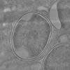 EMDB-16417: Tomogram of an intracellular Salmonella within a ruptured SCV  EMDB-16418: Tomogram of ruptured SCV with early phagophores 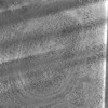 EMDB-16419: Tomogram of stick-shaped contacts between an extended phagophore and the ER around SCV 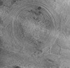 EMDB-16420: Tomogram of three extended phagophores around SCV 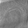 EMDB-16421: Tomogram of omegasomes around SCV 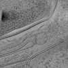 EMDB-16422: Tomogram of membrane contacts between a dilated phagophore rim and the ER around SCV |
| Source |
|
 Homo sapiens (human)