 Movie
Movie Controller
 Structure viewers
 About Yorodumi Papers


+Search query
-Structure paper
| Title | Visualising the cytoskeletal machinery in neuronal growth cones using cryo-electron tomography. |
|---|---|
| Journal, issue, pages | J Cell Sci, Vol. 135, Issue 7, Year 2022 |
| Publish date | Apr 1, 2022 |
 Authors Authors | Joseph Atherton / Melissa Stouffer / Fiona Francis / Carolyn A Moores /    |
| PubMed Abstract | Neurons extend axons to form the complex circuitry of the mature brain. This depends on the coordinated response and continuous remodelling of the microtubule and F-actin networks in the axonal ...Neurons extend axons to form the complex circuitry of the mature brain. This depends on the coordinated response and continuous remodelling of the microtubule and F-actin networks in the axonal growth cone. Growth cone architecture remains poorly understood at nanoscales. We therefore investigated mouse hippocampal neuron growth cones using cryo-electron tomography to directly visualise their three-dimensional subcellular architecture with molecular detail. Our data showed that the hexagonal arrays of actin bundles that form filopodia penetrate and terminate deep within the growth cone interior. We directly observed the modulation of these and other growth cone actin bundles by alteration of individual F-actin helical structures. Microtubules with blunt, slightly flared or gently curved ends predominated in the growth cone, frequently contained lumenal particles and exhibited lattice defects. Investigation of the effect of absence of doublecortin, a neurodevelopmental cytoskeleton regulator, on growth cone cytoskeleton showed no major anomalies in overall growth cone organisation or in F-actin subpopulations. However, our data suggested that microtubules sustained more structural defects, highlighting the importance of microtubule integrity during growth cone migration. |
 External links External links | J Cell Sci / PubMed:35383828 / PubMed Central |
| Methods | EM (subtomogram averaging) / EM (tomography) |
| Resolution | 27.0 - 31.0 Å |
| Structure data |  EMDB-14386: In situ subtomogram average of long-repeat F-actin from neuronal growth cones 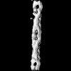 EMDB-14395: In situ subtomogram average of short-repeat F-actin from neuronal growth cones  EMDB-14396: Tomogram of wild-type mouse neuronal growth cone (T-zone), 4 x binned, processed.  EMDB-14397: Tomogram of wild-type mouse neuronal growth cone (P-zone), 4 x binned, processed. 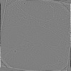 EMDB-14400: Tomogram of wild-type mouse neuronal growth cone (C-zone), 4 x binned, processed.  EMDB-14401: Tomogram of doublecortin knock-out mouse neuronal growth cone (C-zone), 4 x binned, processed.  EMDB-14402: Tomogram of doublecortin knock-out mouse neuronal growth cone (P-zone), 4 x binned, processed. 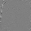 EMDB-14414: Tomogram of doublecortin knock-out mouse neuronal growth cone (T-zone), 4 x binned, processed. 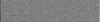 EMDB-14416: Tomogram of wild-type mouse neuronal growth cone (P-zone), 2 x binned, processed. |
| Source |
|

