 Movie
Movie Controller
 Structure viewers
 About Yorodumi Papers


+Search query
-Structure paper
| Title | Architecture of the Tuberous Sclerosis Protein Complex. |
|---|---|
| Journal, issue, pages | J Mol Biol, Vol. 433, Issue 2, Page 166743, Year 2021 |
| Publish date | Jan 22, 2021 |
 Authors Authors | Kailash Ramlaul / Wencheng Fu / Hua Li / Natàlia de Martin Garrido / Lin He / Manjari Trivedi / Wei Cui / Christopher H S Aylett / Geng Wu /   |
| PubMed Abstract | The Tuberous Sclerosis Complex (TSC) protein complex (TSCC), comprising TSC1, TSC2, and TBC1D7, is widely recognised as a key integration hub for cell growth and intracellular stress signals upstream ...The Tuberous Sclerosis Complex (TSC) protein complex (TSCC), comprising TSC1, TSC2, and TBC1D7, is widely recognised as a key integration hub for cell growth and intracellular stress signals upstream of the mammalian target of rapamycin complex 1 (mTORC1). The TSCC negatively regulates mTORC1 by acting as a GTPase-activating protein (GAP) towards the small GTPase Rheb. Both human TSC1 and TSC2 are important tumour suppressors, and mutations in them underlie the disease tuberous sclerosis. We used single-particle cryo-EM to reveal the organisation and architecture of the complete human TSCC. We show that TSCC forms an elongated scorpion-like structure, consisting of a central "body", with a "pincer" and a "tail" at the respective ends. The "body" is composed of a flexible TSC2 HEAT repeat dimer, along the surface of which runs the TSC1 coiled-coil backbone, breaking the symmetry of the dimer. Each end of the body is structurally distinct, representing the N- and C-termini of TSC1; a "pincer" is formed by the highly flexible N-terminal TSC1 core domains and a barbed "tail" makes up the TSC1 coiled-coil-TBC1D7 junction. The TSC2 GAP domain is found abutting the centre of the body on each side of the dimerisation interface, poised to bind a pair of Rheb molecules at a similar separation to the pair in activated mTORC1. Our architectural dissection reveals the mode of association and topology of the complex, casts light on the recruitment of Rheb to the TSCC, and also hints at functional higher order oligomerisation, which has previously been predicted to be important for Rheb-signalling suppression. |
 External links External links | J Mol Biol / PubMed:33307091 / PubMed Central |
| Methods | EM (single particle) |
| Resolution | 4.2 - 8.2 Å |
| Structure data | 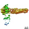 EMDB-11816: 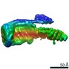 EMDB-11817: 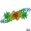 EMDB-11819: |
| Source |
|
 Homo sapiens (human)