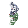
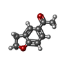
PDB-8i1w: The asymmetric structure of homodimeric E. coli TrpRS bound with tryptophanyl adenylate at one of its two active pockets 手法: X-RAY DIFFRACTION / 解像度: 1.8 Å
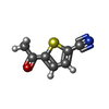
PDB-8i1y: The structure of E. coli TrpRS bound with a chemical fragment 手法: X-RAY DIFFRACTION / 解像度: 1.78 Å
PDB-8i1z: E. coli tryptophanyl-tRNA synthetase bound with a chemical fragment 手法: X-RAY DIFFRACTION / 解像度: 1.8 Å
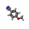
PDB-8i27: E. coli tryptophanyl-tRNA synthetase bound with a chemical fragment at the dimerization interface 手法: X-RAY DIFFRACTION / 解像度: 1.95 Å
PDB-8i2a: E. coli tryptophanyl-tRNA synthetase bound with a chemical fragment at the dimerization interface 手法: X-RAY DIFFRACTION / 解像度: 2.35 Å
PDB-8i2c: E. coli tryptophanyl-tRNA synthetase bound with a chemical fragment at the dimerization interface 手法: X-RAY DIFFRACTION / 解像度: 2.07 Å
PDB-8i2j: E. coli tryptophanyl-tRNA synthetase bound with a chemical fragment at the dimerization interface 手法: X-RAY DIFFRACTION / 解像度: 2.65 Å
PDB-8i2l: E. coli tryptophanyl-tRNA synthetase bound with a chemical fragment at the dimerization interface 手法: X-RAY DIFFRACTION / 解像度: 1.95 Å
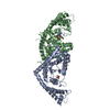
PDB-8i2m: The crystal structure of homodimeric E. coli tryptophanyl-tRNA synthetase bound with niraparib at one of its two active sites 手法: X-RAY DIFFRACTION / 解像度: 2.1 Å
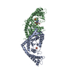
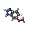
PDB-8i4i: The asymmetric structure of homodimeric E. coli TrpRS bound with tryptophanyl adenylate and L-tryptophan 手法: X-RAY DIFFRACTION / 解像度: 2.2 Å
 ムービー
ムービー コントローラー
 構造ビューア
 万見文献について


 著者
著者 リンク
リンク


 キーワード
キーワード
