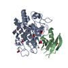
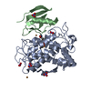
PDB-5z0d: 1.16 A-resolution crystal structure of the deoxy-form tyrosinase from Streptomyces castaneoglobisporus in complex with the caddie protein 手法: X-RAY DIFFRACTION / 解像度: 1.16 Å
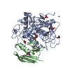
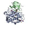
PDB-5z0e: Crystal structure of copper-bound tyrosinase from Streptomyces castaneoglobisporus in complex with the Y98F mutant of the caddie protein obtained by soaking in the hydroxylamine-containing solution for 2 h at 298 K 手法: X-RAY DIFFRACTION / 解像度: 1.16 Å
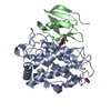
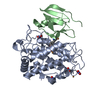
PDB-5z0f: Crystal structure of copper-bound tyrosinase from Streptomyces castaneoglobisporus in complex with the caddie protein obtained by soaking in the hydroxylamine-containing solution for 10 min at 298 K 手法: X-RAY DIFFRACTION / 解像度: 1.16 Å
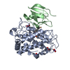
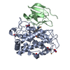
PDB-5z0g: Crystal structure of copper-bound tyrosinase from Streptomyces castaneoglobisporus in complex with the caddie protein obtained by soaking in the hydroxylamine-containing solution for 20 min at 298 K 手法: X-RAY DIFFRACTION / 解像度: 1.32 Å
PDB-5z0h: Crystal structure of copper-bound tyrosinase from Streptomyces castaneoglobisporus in complex with the caddie protein obtained by soaking in the hydroxylamine-containing solution for 2 h at 298 K 手法: X-RAY DIFFRACTION / 解像度: 1.18 Å
PDB-5z0i: Crystal structure of copper-bound tyrosinase from Streptomyces castaneoglobisporus in complex with the caddie protein obtained by soaking in the hydroxylamine-containing solution for 1 h at 277 K 手法: X-RAY DIFFRACTION / 解像度: 1.32 Å
PDB-5z0j: Crystal structure of copper-bound tyrosinase from Streptomyces castaneoglobisporus in complex with the caddie protein obtained by soaking in the hydroxylamine-containing solution for 2 h at 277 K 手法: X-RAY DIFFRACTION / 解像度: 1.35 Å
PDB-5z0k: Crystal structure of copper-bound tyrosinase from Streptomyces castaneoglobisporus in complex with the caddie protein obtained by soaking in the hydroxylamine-containing solution for 4 h at 277 K 手法: X-RAY DIFFRACTION / 解像度: 1.28 Å
PDB-5z0l: Crystal structure of copper-bound tyrosinase from Streptomyces castaneoglobisporus in complex with the caddie protein obtained by soaking in the hydroxylamine-containing solution for 9 h at 277 K 手法: X-RAY DIFFRACTION / 解像度: 1.17 Å
PDB-5z0m: Crystal structure of copper-bound H63F-mutated tyrosinase from Streptomyces castaneoglobisporus in complex with the caddie protein obtained by soaking in the hydroxylamine-containing solution for 12 h at 298 K 手法: X-RAY DIFFRACTION / 解像度: 1.7 Å
 ムービー
ムービー コントローラー
 構造ビューア
 万見文献について


 著者
著者 リンク
リンク


 キーワード
キーワード streptomyces castaneoglobisporus (バクテリア)