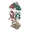
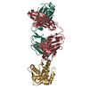
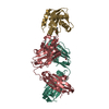
Lipid interactions and angle of approach to the HIV-1 viral membrane of broadly neutralizing antibody 10E8: Insights for vaccine and therapeutic design.
PDB-5sy8: Crystal structure of the complex of 10E8 Fab light chain mutant1 and T117v2 HIV-1 MPER scaffold 手法: X-RAY DIFFRACTION / 解像度: 1.62 Å
PDB-5t29: Crystal structure of 10E8 Fab light chain mutant3, against the MPER region of the HIV-1 Env, in complex with the MPER epitope scaffold T117v2 手法: X-RAY DIFFRACTION / 解像度: 2.03 Å
PDB-5t5b: CRYSTAL STRUCTURE OF THE COMPLEX OF 10E8 FAB LIGHT CHAIN MUTANT5 AND T117V2 HIV-1 MPER SCAFFOLD 手法: X-RAY DIFFRACTION / 解像度: 2.07 Å
PDB-5t6l: Crystal structure of 10E8 Fab in complex with the MPER epitope scaffold T117v2 手法: X-RAY DIFFRACTION / 解像度: 2.1 Å
PDB-5t80: Crystal structure of 10E8 Fab in complex with the MPER epitope scaffold T117v2 and phosphatidic acid (06:0 PA) 手法: X-RAY DIFFRACTION / 解像度: 2.62 Å
PDB-5t85: Crystal structure of 10E8 Fab in complex with the MPER epitope scaffold T117v2 and phosphatidylglycerol (06:0 PG) 手法: X-RAY DIFFRACTION / 解像度: 2.373 Å
PDB-5tfw: Crystal structure of 10E8 Fab light chain mutant2 against the MPER region of the HIV-1 Env, in complex with T117v2 epitope scaffold 手法: X-RAY DIFFRACTION / 解像度: 2.168 Å
 ムービー
ムービー コントローラー
 構造ビューア
 万見文献について


 著者
著者 リンク
リンク


 キーワード
キーワード homo sapiens (ヒト)