| タイトル | Pushing the Limits of Detection of Weak Binding Using Fragment-Based Drug Discovery: Identification of New Cyclophilin Binders. |
|---|
| ジャーナル・号・ページ | J. Mol. Biol., Vol. 429, Page 2556-2570, Year 2017 |
|---|
| 掲載日 | 2017年4月13日 (構造データの登録日) |
|---|
 著者 著者 | Georgiou, C. / McNae, I. / Wear, M. / Ioannidis, H. / Michel, J. / Walkinshaw, M. |
|---|
 リンク リンク |  J. Mol. Biol. / J. Mol. Biol. /  PubMed:28673552 PubMed:28673552 |
|---|
| 手法 | X線回折 |
|---|
| 解像度 | 1.3 - 2 Å |
|---|
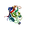
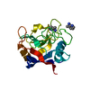
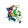
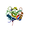
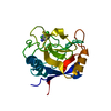
| 構造データ | PDB-5noq:
Structure of cyclophilin A in complex with 3-chloropyridin-2-amine
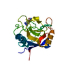
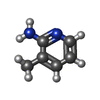
手法: X-RAY DIFFRACTION / 解像度: 1.6 Å PDB-5nor:
Structure of cyclophilin A in complex with 3-methylpyridin-2-amine
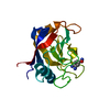
手法: X-RAY DIFFRACTION / 解像度: 1.8 Å PDB-5nos:
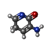
Structure of cyclophilin A in complex with 3-amino-1H-pyridin-2-one
手法: X-RAY DIFFRACTION / 解像度: 1.35 Å PDB-5not:
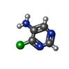
Structure of cyclophilin A in complex with 4-chloropyrimidin-5-amine
手法: X-RAY DIFFRACTION / 解像度: 1.45 Å PDB-5nou:
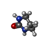
Structure of cyclophilin A in complex with hexahydropyrimidin-2-one
手法: X-RAY DIFFRACTION / 解像度: 1.3 Å PDB-5nov:
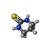
Structure of cyclophilin A in complex with hexahydropyrimidine-2-thione
手法: X-RAY DIFFRACTION / 解像度: 2 Å PDB-5now:
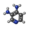
Structure of cyclophilin A in complex with pyridine-3,4-diamine
手法: X-RAY DIFFRACTION / 解像度: 1.48 Å PDB-5nox:
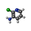
Structure of cyclophilin A in complex with 2-chloropyridin-3-amine
手法: X-RAY DIFFRACTION / 解像度: 1.49 Å PDB-5noy:
Structure of cyclophilin A in complex with 3,4-diaminobenzamide
手法: X-RAY DIFFRACTION / 解像度: 1.43 Å PDB-5noz:
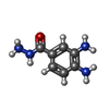
Structure of cyclophilin A in complex with 3,4-diaminobenzohydrazide
手法: X-RAY DIFFRACTION / 解像度: 1.61 Å |
|---|
| 化合物 | |
|---|
| 由来 |  homo sapiens (ヒト) homo sapiens (ヒト)
|
|---|
 キーワード キーワード | ISOMERASE / LIGAND COMPLEX / BETA BARREL / PROLYL CIS/TRANS ISOMERASE / CYTOSOLIC |
|---|
 著者
著者 リンク
リンク J. Mol. Biol. /
  PubMed:28673552






 キーワード
キーワード ムービー
ムービー コントローラー
 構造ビューア
 万見文献について


 homo sapiens (ヒト)