 ムービー
ムービー コントローラー
 構造ビューア
 万見文献について


+検索条件
-Structure paper
| タイトル | An additional substrate binding site in a bacterial phenylalanine hydroxylase. |
|---|---|
| ジャーナル・号・ページ | Eur. Biophys. J., Vol. 42, Page 691-708, Year 2013 |
| 掲載日 | 2011年8月9日 (構造データの登録日) |
 著者 著者 | Ronau, J.A. / Paul, L.N. / Fuchs, J.E. / Corn, I.R. / Wagner, K.T. / Liedl, K.R. / Abu-Omar, M.M. / Das, C. |
 リンク リンク | Eur. Biophys. J. / PubMed:23860686 |
| 手法 | X線回折 |
| 解像度 | 1.35 - 2.13 Å |
| 構造データ |  PDB-3tcy: 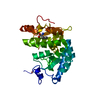 PDB-3tk2: 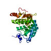 PDB-3tk4:  PDB-4esm: 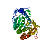 PDB-4etl:  PDB-4jpx: 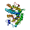 PDB-4jpy: |
| 化合物 |  ChemComp-PHE:  ChemComp-CO:  ChemComp-EDO:  ChemComp-HOH:  ChemComp-FE: |
| 由来 |
|
 キーワード キーワード | OXIDOREDUCTASE / PHENYLALANINE HYDROXYLASE / SUBSTRATE-PROTEIN COMPLEX / DISTAL SITE / phenylalanine bound structure / Protein-Substate complex / Mixed alpha / beta / Hydroxylase / Phenylalanine / Tetrahydrobiopterin / Iron (II) / Molecular oxygen / Hydroxylation / Cobalt-bound structure / Mixed alpha beta / Mutation / Alpha helix-Beta sheet / 5 / 6 / 7 / 8-tetrahydrobiopterin / Fe / Mixed alpha helix-beta sheet / PKU |
 chromobacterium violaceum (バクテリア)