Gökhan Tolun / Camasamudram Vijayasarathy / Rick Huang / Yong Zeng / Yan Li / Alasdair C Steven / Paul A Sieving / J Bernard Heymann /
PubMed 要旨
Retinoschisin (RS1) is involved in cell-cell junctions in the retina, but is unique among known cell-adhesion proteins in that it is a soluble secreted protein. Loss-of-function mutations in RS1 lead ...Retinoschisin (RS1) is involved in cell-cell junctions in the retina, but is unique among known cell-adhesion proteins in that it is a soluble secreted protein. Loss-of-function mutations in RS1 lead to early vision impairment in young males, called X-linked retinoschisis. The disease is characterized by separation of inner retinal layers and disruption of synaptic signaling. Using cryo-electron microscopy, we report the structure at 4.1 Å, revealing double octamer rings not observed before. Each subunit is composed of a discoidin domain and a small N-terminal (RS1) domain. The RS1 domains occupy the centers of the rings, but are not required for ring formation and are less clearly defined, suggesting mobility. We determined the structure of the discoidin rings, consistent with known intramolecular and intermolecular disulfides. The interfaces internal to and between rings feature residues implicated in X-linked retinoschisis, indicating the importance of correct assembly. Based on this structure, we propose that RS1 couples neighboring membranes together through octamer-octamer contacts, perhaps modulated by interactions with other membrane components.
EMDB-6425: Retinoschisin, back-to-back octameric rings PDB-3jd6: Double octamer structure of retinoschisin, a cell-cell adhesion protein of the retina 手法: EM (単粒子) / 解像度: 4.1 Å
 ムービー
ムービー コントローラー
 構造ビューア
 万見文献について


 著者
著者
 リンク
リンク キーワード
キーワード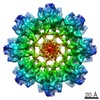

 homo sapiens (ヒト)