 ムービー
ムービー コントローラー
 構造ビューア
 万見文献について


+検索条件
-Structure paper
| タイトル | Biochemical and Structural Characterization of Residue 96 Mutants of Plasmodium Falciparum Triosephosphate Isomerase: Active-Site Loop Conformation, Hydration and Identification of a Dimer-Interface Ligand-Binding Site. |
|---|---|
| ジャーナル・号・ページ | Acta Crystallogr. ,Sect. D, Vol. 65, Page 847-, Year 2009 |
| 掲載日 | 2007年11月3日 (構造データの登録日) |
 著者 著者 | Gayathri, P. / Banerjee, M. / Vijayalakshmi, A. / Balaram, H. / Balaram, P. / Murthy, M.R.N. |
 リンク リンク | Acta Crystallogr. ,Sect. D / PubMed:19622869 |
| 手法 | X線回折 |
| 解像度 | 1.4 - 2.25 Å |
| 構造データ | 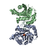 PDB-2vfd: 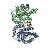 PDB-2vfe:  PDB-2vff: 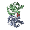 PDB-2vfg: 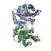 PDB-2vfh: 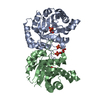 PDB-2vfi: |
| 化合物 |  ChemComp-SO4:  ChemComp-HOH:  ChemComp-3PG:  ChemComp-GOL: |
| 由来 |
|
 キーワード キーワード | ISOMERASE / PLASMODIUM FALCIPARUM / FATTY ACID BIOSYNTHESIS / TRIOSEPHOSPHATE ISOMERASE / PENTOSE SHUNT / GLUCONEOGENESIS / LIPID SYNTHESIS / TIM / MUTANT / LOOP OPEN / GLYCOLYSIS / 3-PHOSPHOGLYCERATE / DIMER INTERFACE / LOOP CLOSED |

