PDB-2ptw: Crystal Structure of the T. brucei enolase complexed with sulphate, identification of a metal binding site IV 手法: X-RAY DIFFRACTION / 解像度: 1.9 Å
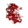
PDB-2ptx: Crystal Structure of the T. brucei enolase complexed with sulphate in closed conformation 手法: X-RAY DIFFRACTION / 解像度: 1.9 Å
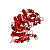
PDB-2pty: Crystal Structure of the T. brucei enolase complexed with PEP 手法: X-RAY DIFFRACTION / 解像度: 2.0 Å
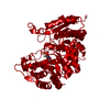
PDB-2ptz: Crystal Structure of the T. brucei enolase complexed with phosphonoacetohydroxamate (PAH), His156-out conformation 手法: X-RAY DIFFRACTION / 解像度: 1.65 Å
PDB-2pu0: Crystal Structure of the T. brucei enolase complexed with phosphonoacetohydroxamate (PAH), His156-in conformation 手法: X-RAY DIFFRACTION / 解像度: 1.9 Å
PDB-2pu1: Crystal Structure of the T. brucei enolase complexed with Fluoro-phosphonoacetohydroxamate (FPAH) 手法: X-RAY DIFFRACTION / 解像度: 1.8 Å
 ムービー
ムービー コントローラー
 構造ビューア
 万見文献について


 著者
著者 リンク
リンク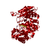


 キーワード
キーワード
