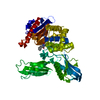
PDB-2ex2: Crystal structure of penicillin binding protein 4 (dacB) from Escherichia coli 手法: X-RAY DIFFRACTION / 解像度: 1.55 Å
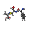
PDB-2ex6: Crystal structure of penicillin binding protein 4 (dacB) from Escherichia coli, complexed with ampicillin 手法: X-RAY DIFFRACTION / 解像度: 1.6 Å
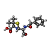
PDB-2ex8: Crystal structure of penicillin binding protein 4 (dacB) from Escherichia coli, complexed with penicillin-G 手法: X-RAY DIFFRACTION / 解像度: 1.6 Å
PDB-2ex9: Crystal structure of penicillin binding protein 4 (dacB) from Escherichia coli, complexed with penicillin-V 手法: X-RAY DIFFRACTION / 解像度: 1.65 Å
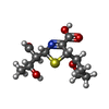
PDB-2exa: Crystal structure of penicillin binding protein 4 (dacB) from Escherichia coli, complexed with FAROM 手法: X-RAY DIFFRACTION / 解像度: 1.7 Å
PDB-2exb: Crystal structure of penicillin binding protein 4 (dacB) from Escherichia coli, complexed with FLOMOX 手法: X-RAY DIFFRACTION / 解像度: 1.75 Å
 ムービー
ムービー コントローラー
 構造ビューア
 万見文献について


 著者
著者 リンク
リンク


 キーワード
キーワード
