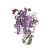
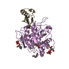
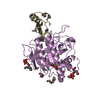
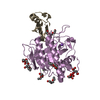
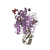
タイトル Binding, Proteolytic, and Crystallographic Analyses of Mutations at the Protease-Inhibitor Interface of the Subtilisin BPN'/Chymotrypsin Inhibitor 2 Complex(,). ジャーナル・号・ページ Biochemistry , Vol. 43, Page 13648-13656, Year 2004掲載日 2004年6月10日 (構造データの登録日) Radisky, E.S. / Kwan, G. / Karen Lu, C.J. / Koshland Jr., D.E. / 手法 X線回折 解像度 1.3 - 1.7 Å 構造データ PDB-1tm1 2 手法 : X-RAY DIFFRACTION / 解像度 : 1.7 Å
PDB-1tm3 M59k mutant手法 : X-RAY DIFFRACTION / 解像度 : 1.57 Å
PDB-1tm4 subtilsin BPN'with chymotrypsin inhibitor 2 M59G mutant手法 : X-RAY DIFFRACTION / 解像度 : 1.7 Å
PDB-1tm5 M59A mutant手法 : X-RAY DIFFRACTION / 解像度 : 1.45 Å
PDB-1tm7 M59Y mutant手法 : X-RAY DIFFRACTION / 解像度 : 1.59 Å
PDB-1tmg M59F mutant手法 : X-RAY DIFFRACTION / 解像度 : 1.67 Å
PDB-1to1 Y61A mutant手法 : X-RAY DIFFRACTION / 解像度 : 1.68 Å
PDB-1to2 in pH 9 cryosoak 手法 : X-RAY DIFFRACTION / 解像度 : 1.3 Å
化合物 由来 bacillus amyloliquefaciens (バクテリア)hordeum vulgare subsp. vulgare (オオムギ) / / / /
 ムービー
ムービー コントローラー
 構造ビューア
 万見文献について


 著者
著者 リンク
リンク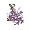


 キーワード
キーワード

