ムービー
ムービー コントローラー
 構造ビューア
 万見文献について


+検索条件
-Structure paper
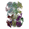
| タイトル | Cryo-EM structure of Mcm2-7 double hexamer on DNA suggests a lagging-strand DNA extrusion model. |
|---|---|
| ジャーナル・号・ページ | Proc Natl Acad Sci U S A, Vol. 114, Issue 45, Page E9529-E9538, Year 2017 |
| 掲載日 | 2017年11月7日 |
 著者 著者 | Yasunori Noguchi / Zuanning Yuan / Lin Bai / Sarah Schneider / Gongpu Zhao / Bruce Stillman / Christian Speck / Huilin Li /   |
| PubMed 要旨 | During replication initiation, the core component of the helicase-the Mcm2-7 hexamer-is loaded on origin DNA as a double hexamer (DH). The two ring-shaped hexamers are staggered, leading to a kinked ...During replication initiation, the core component of the helicase-the Mcm2-7 hexamer-is loaded on origin DNA as a double hexamer (DH). The two ring-shaped hexamers are staggered, leading to a kinked axial channel. How the origin DNA interacts with the axial channel is not understood, but the interaction could provide key insights into Mcm2-7 function and regulation. Here, we report the cryo-EM structure of the Mcm2-7 DH on dsDNA and show that the DNA is zigzagged inside the central channel. Several of the Mcm subunit DNA-binding loops, such as the oligosaccharide-oligonucleotide loops, helix 2 insertion loops, and presensor 1 (PS1) loops, are well defined, and many of them interact extensively with the DNA. The PS1 loops of Mcm 3, 4, 6, and 7, but not 2 and 5, engage the lagging strand with an approximate step size of one base per subunit. Staggered coupling of the two opposing hexamers positions the DNA right in front of the two Mcm2-Mcm5 gates, with each strand being pressed against one gate. The architecture suggests that lagging-strand extrusion initiates in the middle of the DH that is composed of the zinc finger domains of both hexamers. To convert the Mcm2-7 DH structure into the Mcm2-7 hexamer structure found in the active helicase, the N-tier ring of the Mcm2-7 hexamer in the DH-dsDNA needs to tilt and shift laterally. We suggest that these N-tier ring movements cause the DNA strand separation and lagging-strand extrusion. |
 リンク リンク | Proc Natl Acad Sci U S A / PubMed:29078375 / PubMed Central |
| 手法 | EM (単粒子) |
| 解像度 | 3.9 Å |
| 構造データ | |
| 化合物 |  ChemComp-ADP: |
| 由来 |
|
 キーワード キーワード | HYDROLASE/DNA / complex / DNA replication / HYDROLASE-DNA complex |