 ムービー
ムービー コントローラー
 構造ビューア
 万見文献について


+検索条件
-Structure paper
| タイトル | Bovine F1Fo ATP synthase monomers bend the lipid bilayer in 2D membrane crystals. |
|---|---|
| ジャーナル・号・ページ | Elife, Vol. 4, Page e06119, Year 2015 |
| 掲載日 | 2015年3月27日 |
 著者 著者 | Chimari Jiko / Karen M Davies / Kyoko Shinzawa-Itoh / Kazutoshi Tani / Shintaro Maeda / Deryck J Mills / Tomitake Tsukihara / Yoshinori Fujiyoshi / Werner Kühlbrandt / Christoph Gerle /   |
| PubMed 要旨 | We have used a combination of electron cryo-tomography, subtomogram averaging, and electron crystallographic image processing to analyse the structure of intact bovine F(1)F(o) ATP synthase in 2D ...We have used a combination of electron cryo-tomography, subtomogram averaging, and electron crystallographic image processing to analyse the structure of intact bovine F(1)F(o) ATP synthase in 2D membrane crystals. ATPase assays and mass spectrometry analysis of the 2D crystals confirmed that the enzyme complex was complete and active. The structure of the matrix-exposed region was determined at 24 Å resolution by subtomogram averaging and repositioned into the tomographic volume to reveal the crystal packing. F(1)F(o) ATP synthase complexes are inclined by 16° relative to the crystal plane, resulting in a zigzag topology of the membrane and indicating that monomeric bovine heart F(1)F(o) ATP synthase by itself is sufficient to deform lipid bilayers. This local membrane curvature is likely to be instrumental in the formation of ATP synthase dimers and dimer rows, and thus for the shaping of mitochondrial cristae. |
 リンク リンク | Elife / PubMed:25815585 / PubMed Central |
| 手法 | EM (サブトモグラム平均) |
| 解像度 | 24.0 Å |
| 構造データ | 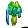 EMDB-2982: |
| 由来 |
|

