 ムービー
ムービー コントローラー
 構造ビューア
 万見文献について


+検索条件
-Structure paper
| タイトル | Electron microscopic imaging revealed the flexible filamentous structure of the cell attachment protein P2 of Rice dwarf virus located around the icosahedral 5-fold axes. |
|---|---|
| ジャーナル・号・ページ | J Biochem, Vol. 159, Issue 2, Page 181-190, Year 2016 |
| 掲載日 | 2015年9月15日 |
 著者 著者 | Naoyuki Miyazaki / Akifumi Higashiura / Tomoko Higashiura / Fusamichi Akita / Hiroyuki Hibino / Toshihiro Omura / Atsushi Nakagawa / Kenji Iwasaki /  |
| PubMed 要旨 | The minor outer capsid protein P2 of Rice dwarf virus (RDV), a member of the genus Phytoreovirus in the family Reoviridae, is essential for viral cell entry. Here, we clarified the structure of P2 ...The minor outer capsid protein P2 of Rice dwarf virus (RDV), a member of the genus Phytoreovirus in the family Reoviridae, is essential for viral cell entry. Here, we clarified the structure of P2 and the interactions to host insect cells. Negative stain electron microscopy (EM) showed that P2 proteins are monomeric and flexible L-shaped filamentous structures of ∼20 nm in length. Cryo-EM structure revealed the spatial arrangement of P2 in the capsid, which was prescribed by the characteristic virion structure. The P2 proteins were visualized as partial rod-shaped structures of ∼10 nm in length in the cryo-EM map and accommodated in crevasses on the viral surface around icosahedral 5-fold axes with hydrophobic interactions. The remaining disordered region of P2 assumed to be extended to the radial direction towards exterior. Electron tomography clearly showed that RDV particles were away from the cellular membrane at a uniform distance and several spike-like densities, probably corresponding to P2, connecting a viral particle to the host cellular membrane during cell entry. By combining the in vitro and in vivo structural information, we could gain new insights into the detailed mechanism of the cell entry of RDV. |
 リンク リンク | J Biochem / PubMed:26374901 / PubMed Central |
| 手法 | EM (単粒子) |
| 解像度 | 13.0 Å |
| 構造データ | 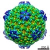 EMDB-2782: |
