 ムービー
ムービー コントローラー
 構造ビューア
 万見文献について


+検索条件
-Structure paper
| タイトル | CryoET shows cofilactin filaments inside the microtubule lumen. |
|---|---|
| ジャーナル・号・ページ | EMBO Rep, Vol. 24, Issue 11, Page e57264, Year 2023 |
| 掲載日 | 2023年11月6日 |
 著者 著者 | Camilla Ventura Santos / Stephen L Rogers / Andrew P Carter /   |
| PubMed 要旨 | Cytoplasmic microtubules are tubular polymers that can harbor small proteins or filaments inside their lumen. The identities of these objects and mechanisms for their accumulation have not been ...Cytoplasmic microtubules are tubular polymers that can harbor small proteins or filaments inside their lumen. The identities of these objects and mechanisms for their accumulation have not been conclusively established. Here, we used cryogenic electron tomography of Drosophila S2 cell protrusions and found filaments inside the microtubule lumen, which resemble those reported recently in human HAP1 cells. The frequency of these filaments increased upon inhibition of the sarco/endoplasmic reticulum Ca ATPase with the small molecule drug thapsigargin. Subtomogram averaging showed that the luminal filaments adopt a helical structure reminiscent of cofilin-bound actin (cofilactin). Consistent with this, we observed cofilin dephosphorylation, an activating modification, in cells under the same conditions that increased luminal filament occurrence. Furthermore, RNA interference knock-down of cofilin reduced the frequency of luminal filaments with cofilactin morphology. These results suggest that cofilin activation stimulates its accumulation on actin filaments inside the microtubule lumen. |
 リンク リンク | EMBO Rep / PubMed:37702953 / PubMed Central |
| 手法 | EM (トモグラフィー) |
| 構造データ | 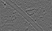 EMDB-18475: Cryo-electron tomogram of an induced S2 cell protrusion. The cell was treated with 2uM thapsigargin (5h) and with 2.5uM Cytochalasin D (2h). |
| 由来 |
|

