 ムービー
ムービー コントローラー
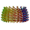
 構造ビューア
 万見文献について


+検索条件
-Structure paper
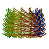
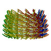
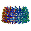
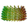
| タイトル | Structural basis for VIPP1 oligomerization and maintenance of thylakoid membrane integrity. |
|---|---|
| ジャーナル・号・ページ | Cell, Vol. 184, Issue 14, Page 3643-3659.e23, Year 2021 |
| 掲載日 | 2021年7月8日 |
 著者 著者 | Tilak Kumar Gupta / Sven Klumpe / Karin Gries / Steffen Heinz / Wojciech Wietrzynski / Norikazu Ohnishi / Justus Niemeyer / Benjamin Spaniol / Miroslava Schaffer / Anna Rast / Matthias Ostermeier / Mike Strauss / Jürgen M Plitzko / Wolfgang Baumeister / Till Rudack / Wataru Sakamoto / Jörg Nickelsen / Jan M Schuller / Michael Schroda / Benjamin D Engel /    |
| PubMed 要旨 | Vesicle-inducing protein in plastids 1 (VIPP1) is essential for the biogenesis and maintenance of thylakoid membranes, which transform light into life. However, it is unknown how VIPP1 performs its ...Vesicle-inducing protein in plastids 1 (VIPP1) is essential for the biogenesis and maintenance of thylakoid membranes, which transform light into life. However, it is unknown how VIPP1 performs its vital membrane-remodeling functions. Here, we use cryo-electron microscopy to determine structures of cyanobacterial VIPP1 rings, revealing how VIPP1 monomers flex and interweave to form basket-like assemblies of different symmetries. Three VIPP1 monomers together coordinate a non-canonical nucleotide binding pocket on one end of the ring. Inside the ring's lumen, amphipathic helices from each monomer align to form large hydrophobic columns, enabling VIPP1 to bind and curve membranes. In vivo mutations in these hydrophobic surfaces cause extreme thylakoid swelling under high light, indicating an essential role of VIPP1 lipid binding in resisting stress-induced damage. Using cryo-correlative light and electron microscopy (cryo-CLEM), we observe oligomeric VIPP1 coats encapsulating membrane tubules within the Chlamydomonas chloroplast. Our work provides a structural foundation for understanding how VIPP1 directs thylakoid biogenesis and maintenance. |
 リンク リンク | Cell / PubMed:34166613 |
| 手法 | EM (トモグラフィー) / EM (単粒子) |
| 解像度 | 3.8 - 5.0 Å |
| 構造データ |  EMDB-12324:  EMDB-12325:  EMDB-12326:  EMDB-12327:  EMDB-12328:  EMDB-12329:  EMDB-12330: EMDB-12710, PDB-7o3w: EMDB-12711, PDB-7o3x: EMDB-12712, PDB-7o3y: EMDB-12713, PDB-7o40: EMDB-12714, PDB-7o3z: |
| 化合物 |  ChemComp-ADP: |
| 由来 |
|
 キーワード キーワード | LIPID BINDING PROTEIN / ESCRT-III / Membrane remodelling / nucleotide hydrolysis / photosynthesis / thylakoid biogenesis / stress response |


 Chlamydomonas reinhardtii (クラミドモナス)