 ムービー
ムービー コントローラー
 構造ビューア
 万見文献について


+検索条件
-Structure paper
| タイトル | Electron cryo-microscopy and image reconstruction of adeno-associated virus type 2 empty capsids. |
|---|---|
| ジャーナル・号・ページ | EMBO Rep, Vol. 2, Issue 11, Page 997-991002, Year 2001 |
| 掲載日 | 2002年1月30日 |
 著者 著者 | S Kronenberg / J A Kleinschmidt / B Böttcher /  |
| PubMed 要旨 | Adeno-associated virus type 2 empty capsids are composed of three proteins, VP1, VP2 and VP3, which have relative molecular masses of 87, 72 and 62 kDa, respectively, and differ in their N-terminal ...Adeno-associated virus type 2 empty capsids are composed of three proteins, VP1, VP2 and VP3, which have relative molecular masses of 87, 72 and 62 kDa, respectively, and differ in their N-terminal amino acid sequences. They have a likely molar ratio of 1:1:8 and occupy symmetrical equivalent positions in an icosahedrally arranged protein shell. We have investigated empty capsids of adeno-associated virus type 2 by electron cryo-microscopy and icosahedral image reconstruction. The three-dimensional map at 1.05 nm resolution showed sets of three elongated spikes surrounding the three-fold symmetry axes and narrow empty channels at the five-fold axes. The inside of the capsid superimposed with the previously determined structure of the canine parvovirus (Q. Xie and M.S. Chapman, 1996, J. Mol. Biol., 264, 497-520), whereas the outer surface showed clear discrepancies. Globular structures at the inner surface of the capsid at the two-fold symmetry axes were identified as possible positions for the N-terminal extensions of VP1 and VP2. |
 リンク リンク | EMBO Rep / PubMed:11713191 / PubMed Central |
| 手法 | EM (単粒子) |
| 解像度 | 10.5 Å |
| 構造データ | 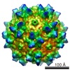 EMDB-1907: |
