 Movie
Movie Controller
 Structure viewers
 About Yorodumi Papers


+Search query
-Structure paper
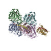
| Title | Cryo-EM structure of VASH1-SVBP bound to microtubules. |
|---|---|
| Journal, issue, pages | Elife, Vol. 9, Year 2020 |
| Publish date | Aug 10, 2020 |
 Authors Authors | Faxiang Li / Yang Li / Xuecheng Ye / Haishan Gao / Zhubing Shi / Xuelian Luo / Luke M Rice / Hongtao Yu /   |
| PubMed Abstract | The dynamic tyrosination-detyrosination cycle of α-tubulin regulates microtubule functions. Perturbation of this cycle impairs mitosis, neural physiology, and cardiomyocyte contraction. The ...The dynamic tyrosination-detyrosination cycle of α-tubulin regulates microtubule functions. Perturbation of this cycle impairs mitosis, neural physiology, and cardiomyocyte contraction. The carboxypeptidases vasohibins 1 and 2 (VASH1 and VASH2), in complex with the small vasohibin-binding protein (SVBP), mediate α-tubulin detyrosination. These enzymes detyrosinate microtubules more efficiently than soluble αβ-tubulin heterodimers. The structural basis for this substrate preference is not understood. Using cryo-electron microscopy (cryo-EM), we have determined the structure of human VASH1-SVBP bound to microtubules. The acidic C-terminal tail of α-tubulin binds to a positively charged groove near the active site of VASH1. VASH1 forms multiple additional contacts with the globular domain of α-tubulin, including contacts with a second α-tubulin in an adjacent protofilament. Simultaneous engagement of two protofilaments by VASH1 can only occur within the microtubule lattice, but not with free αβ heterodimers. These lattice-specific interactions enable preferential detyrosination of microtubules by VASH1. |
 External links External links | Elife / PubMed:32773040 / PubMed Central |
| Methods | EM (single particle) |
| Resolution | 3.1 Å |
| Structure data | EMDB-21893, PDB-6wsl: |
| Chemicals |  ChemComp-GTP:  ChemComp-G2P: |
| Source |
|
 Keywords Keywords | PROTEIN BINDING / Microtubule / Posttranslational modification / Detyrosination / Vasohibin |
 homo sapiens (human)