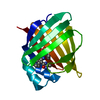
PDB-7fvz: Crystal Structure of human FABP4 in complex with (E)-6-(5-methoxy-3,6,7-trimethyl-1,2-benzoxazol-4-yl)-4-methyl-hex-4-enoic acid Method: X-RAY DIFFRACTION / Resolution: 1.12 Å
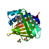
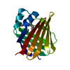
PDB-7fwf: Crystal Structure of human FABP4 binding site mutated to that of FABP5 in complex with 5-cyclohexyl-6-(2,2,2-trifluoroethoxy)-2-(trifluoromethyl)pyridine-3-carboxylic acid, i.e. SMILES c1(c(nc(c(c1)C(=O)O)C(F)(F)F)OCC(F)(F)F)C1CCCCC1 with IC50=0.733066 microM Method: X-RAY DIFFRACTION / Resolution: 1.24 Å
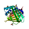
PDB-7fwv: Crystal Structure of human FABP4 binding site mutated to that of FABP5 in complex with 6-cyclopentyl-N,5-dimethyl-4-phenyl-N-propan-2-yl-3-(1H-tetrazol-5-yl)pyridin-2-amine, i.e. SMILES c1(c(nc(c(c1c1ccccc1)C1=NN=NN1)N(C(C)C)C)C1CCCC1)C with IC50=0.0609504 microM Method: X-RAY DIFFRACTION / Resolution: 1.45 Å
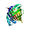
PDB-7fwx: Crystal Structure of human FABP4 binding site mutated to that of FABP5 Method: X-RAY DIFFRACTION / Resolution: 1.13 Å
PDB-7fx2: Crystal Structure of human FABP4 binding site mutated to that of FABP5 in complex with 2-[[6,6-difluoro-3-(4-methyl-1,3-thiazol-2-yl)-5,7-dihydro-4H-1-benzothiophen-2-yl]carbamoyl]cyclohexene-1-carboxylic acid, i.e. SMILES N(C(=O)C1=C(CCCC1)C(=O)O)C1=C(C2=NC(=CS2)C)C2=C(S1)CC(CC2)(F)F with IC50=0.0587459 microM Method: X-RAY DIFFRACTION / Resolution: 1.47 Å
PDB-7fxl: Crystal Structure of human FABP4 binding site mutated to that of FABP5 in complex with myristic acid Method: X-RAY DIFFRACTION / Resolution: 1.12 Å
PDB-7fxs: Crystal Structure of human FABP4 binding site mutated to that of FABP5 in complex with 5-phenoxy-6-(2,2,2-trifluoroethoxy)-2-(trifluoromethyl)pyridine-3-carboxylic acid, i.e. SMILES c1(c(nc(c(c1)C(=O)O)C(F)(F)F)OCC(F)(F)F)Oc1ccccc1 with IC50=2.33933 microM Method: X-RAY DIFFRACTION / Resolution: 1.25 Å
PDB-7fy4: Crystal Structure of human FABP4 binding site mutated to that of FABP5 in complex with 2-benzyl-6-tert-butyl-3-methyl-4-phenyl-5-(1H-tetrazol-5-yl)pyridine, i.e. SMILES N1C(=NN=N1)c1c(nc(c(c1c1ccccc1)C)Cc1ccccc1)C(C)(C)C with IC50=0.0608835 microM Method: X-RAY DIFFRACTION / Resolution: 1.51 Å
PDB-7fy9: Crystal Structure of human FABP4 binding site mutated to that of FABP5 in complex with 2-cyclopentyl-4-(4-fluorophenyl)-6-[1-(methoxymethyl)cyclopentyl]-3-methyl-5-(1H-tetrazol-5-yl)pyridine, i.e. SMILES c1(c(nc(c(c1c1ccc(cc1)F)C1=NN=NN1)C1(CCCC1)COC)C1CCCC1)C with IC50=0.14164 microM Method: X-RAY DIFFRACTION / Resolution: 1.74 Å
PDB-7fyh: Crystal Structure of human FABP4 binding site mutated to that of FABP5 in complex with 6-fluoro-1,3-benzothiazol-2-amine Method: X-RAY DIFFRACTION / Resolution: 1.34 Å
PDB-7fym: Crystal Structure of human FABP4 binding site mutated to that of FABP5 in complex with 5-(3-bromo-4-methylphenyl)-3,3-dimethyl-5-oxopentanoic acid, i.e. SMILES c1(C(=O)CC(CC(=O)O)(C)C)cc(c(cc1)C)Br with IC50=5.1 microM Method: X-RAY DIFFRACTION / Resolution: 1.21 Å
PDB-7fzt: Crystal Structure of human FABP4 binding site mutated to that of FABP5 in complex with rac-(1R,2S)-2-[(3,4-dichlorobenzoyl)amino]cyclohexane-1-carboxylic acid, i.e. SMILES C1CC[C@@H]([C@@H](C1)C(=O)O)NC(=O)c1cc(c(cc1)Cl)Cl with IC50=15.8182 microM Method: X-RAY DIFFRACTION / Resolution: 1.4 Å
PDB-7fzu: Crystal Structure of human FABP4 binding site mutated to that of FABP5 in complex with 2-(indole-1-carbonylamino)benzoic acid, i.e. SMILES c12N(C(=O)Nc3c(cccc3)C(=O)O)C=Cc1cccc2 with IC50=18.1696 microM Method: X-RAY DIFFRACTION / Resolution: 1.25 Å
PDB-7g0c: Crystal Structure of human FABP4 binding site mutated to that of FABP5 in complex with 2-[2,3-bis[(2-chlorophenyl)methoxy]phenyl]-2-methoxyacetic acid, i.e. SMILES c1c(c(c(cc1)OCc1ccccc1Cl)OCc1c(cccc1)Cl)[C@@H](C(=O)O)OC with IC50=1.1 microM Method: X-RAY DIFFRACTION / Resolution: 1.14 Å
PDB-7g0o: Crystal Structure of human FABP4 binding site mutated to that of FABP5 in complex with 2-[[3-(3-cyclopropyl-1,2,4-oxadiazol-5-yl)-4,5-dimethylthiophen-2-yl]carbamoyl]cyclohexene-1-carboxylic acid, i.e. SMILES C1(=C(CCCC1)C(=O)NC1=C(C(=C(C)S1)C)C1=NC(=NO1)C1CC1)C(=O)O with IC50=0.0950978 microM Method: X-RAY DIFFRACTION / Resolution: 1.32 Å
PDB-7g0x: Crystal Structure of human FABP4 binding site mutated to that of FABP5 in complex with isoquinolin-3-amine Method: X-RAY DIFFRACTION / Resolution: 1.48 Å
PDB-7g0z: Crystal Structure of human FABP4 binding site mutated to that of FABP5 in complex with 5-[[3-(3-cyclopropyl-1,2,4-oxadiazol-5-yl)-4,5,6,7-tetrahydro-1-benzothiophen-2-yl]carbamoyl]-3,6-dihydro-2H-pyran-4-carboxylic acid, i.e. SMILES S1C(=C(C2=NC(=NO2)C2CC2)C2=C1CCCC2)NC(=O)C1=C(CCOC1)C(=O)O with IC50=0.48537 microM Method: X-RAY DIFFRACTION / Resolution: 0.84 Å
PDB-7g12: Crystal Structure of human FABP4 binding site mutated to that of FABP5 in complex with N-methyl-6-(3-methylthiophen-2-yl)-4-phenyl-N-propan-2-yl-3-(1H-tetrazol-5-yl)pyridin-2-amine, i.e. SMILES c1c(nc(c(c1c1ccccc1)C1=NN=NN1)N(C(C)C)C)C1=C(C=CS1)C with IC50=2.97112 microM Method: X-RAY DIFFRACTION / Resolution: 1.64 Å
PDB-7g13: Crystal Structure of human FABP4 binding site mutated to that of FABP5 in complex with 4-[[3-(3-cyclopropyl-1,2,4-oxadiazol-5-yl)-4,5,6,7-tetrahydro-1-benzothiophen-2-yl]carbamoyl]-3,6-dihydro-2H-pyran-5-carboxylic acid, i.e. SMILES S1C(=C(C2=NC(=NO2)C2CC2)C2=C1CCCC2)NC(=O)C1=C(COCC1)C(=O)O with IC50=0.119472 microM Method: X-RAY DIFFRACTION / Resolution: 1.15 Å
PDB-7g15: Crystal Structure of human FABP4 binding site mutated to that of FABP5 in complex with rac-(1R,2S)-2-[(3,4-dichlorophenoxy)methyl]cyclohexane-1-carboxylic acid, i.e. SMILES C1CC[C@H]([C@H](C1)C(=O)O)COc1cc(c(cc1)Cl)Cl with IC50=6.03742 microM Method: X-RAY DIFFRACTION / Resolution: 1.28 Å
PDB-7g1m: Crystal Structure of human FABP4 binding site mutated to that of FABP5 in complex with rac-(1R,2R)-2-[[3-(3-methyl-1,2,4-oxadiazol-5-yl)-4,5,6,7-tetrahydro-1-benzothiophen-2-yl]carbamoyl]cyclohexane-1-carboxylic acid, i.e. SMILES C1(=C(C2=C(S1)CCCC2)C1=NC(=NO1)C)NC(=O)[C@@H]1[C@H](CCCC1)C(=O)O with IC50=0.365 microM Method: X-RAY DIFFRACTION / Resolution: 1.34 Å
PDB-7g1p: Crystal Structure of human FABP4 binding site mutated to that of FABP5 in complex with 6-chloro-5-fluoro-1H-benzimidazole Method: X-RAY DIFFRACTION / Resolution: 1.28 Å
PDB-7g1w: Crystal Structure of human FABP4 binding site mutated to that of FABP5 in complex with 5-(3,5-dichloroanilino)-3,3-dimethyl-5-oxopentanoic acid, i.e. SMILES C(=O)(Nc1cc(cc(c1)Cl)Cl)CC(CC(=O)O)(C)C with IC50=4.4 microM Method: X-RAY DIFFRACTION / Resolution: 1.34 Å
In the structure databanks used in Yorodumi, some data are registered as the other names, "COVID-19 virus" and "2019-nCoV". Here are the details of the virus and the list of structure data.
Jan 31, 2019. EMDB accession codes are about to change! (news from PDBe EMDB page)
EMDB accession codes are about to change! (news from PDBe EMDB page)
The allocation of 4 digits for EMDB accession codes will soon come to an end. Whilst these codes will remain in use, new EMDB accession codes will include an additional digit and will expand incrementally as the available range of codes is exhausted. The current 4-digit format prefixed with “EMD-” (i.e. EMD-XXXX) will advance to a 5-digit format (i.e. EMD-XXXXX), and so on. It is currently estimated that the 4-digit codes will be depleted around Spring 2019, at which point the 5-digit format will come into force.
The EM Navigator/Yorodumi systems omit the EMD- prefix.
Related info.:Q: What is EMD? / ID/Accession-code notation in Yorodumi/EM Navigator
 Movie
Movie Controller
 Structure viewers
 About Yorodumi Papers


 Authors
 External links
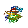


 Keywords
 homo sapiens (human)