 Movie
Movie Controller
 Structure viewers
 About Yorodumi Papers


+Search query
-Structure paper
| Title | A disulfide redox switch mechanism regulates glycoside hydrolase function. |
|---|---|
| Journal, issue, pages | Nat Commun, Vol. 17, Issue 1, Page 45, Year 2026 |
| Publish date | Jan 5, 2026 |
 Authors Authors | Marcele Pandeló Martins / Gustavo Henrique Martins / Felipe Jun Fuzita / João Paulo Menezes Spadeto / Renan Yuji Miyamoto / Felippe Mariano Colombari / Fabiane Stoffel / Luciano Graciani Dolce / Camila Ramos Dos Santos / Rodrigo Silva Araujo Streit / Antônio Carlos Borges / Rafael Henrique Galinari / Yoshihisa Yoshimi / Paul Dupree / Gabriela Felix Persinoti / Mariana Abrahão Bueno Morais / Mario Tyago Murakami /    |
| PubMed Abstract | Disulfide bonds are a key post-translational modification involved in protein folding, structural stability, and functional regulation. Here, we demonstrate that a glycoside hydrolase from the GH2 ...Disulfide bonds are a key post-translational modification involved in protein folding, structural stability, and functional regulation. Here, we demonstrate that a glycoside hydrolase from the GH2 family undergoes reversible redox regulation through an intramolecular disulfide bond. The enzyme is inactive in its oxidized state and becomes active when reduced through a fully reversible process. Under oxidative conditions, multiple crystallographic and cryo-EM structures revealed a pronounced structural disorder in the active site, most prominent in the regulatory and catalytic loops, which disrupts the substrate binding site and, remarkably, the configuration of the acidic catalytic residues. Conversely, a high-resolution cryo-EM structure of the active (reduced) state unveiled a well-ordered active site with catalytic residues properly positioned for a classical Koshland retaining mechanism. This reversible order-disorder process based on a disulfide switch provides a mechanism for redox-dependent control of glycoside hydrolase activity, with potential implications for carbohydrate metabolism, microbial adaptation and biotechnological applications. |
 External links External links | Nat Commun / PubMed:41491304 / PubMed Central |
| Methods | EM (single particle) / X-ray diffraction |
| Resolution | 2 - 3.41 Å |
| Structure data |  EMDB-49363: Cryo-EM map of the inactive conformation of a glycoside hydrolase (CapGH2b) from the GH2 family EMDB-49364, PDB-9nfe:  PDB-9np8:  PDB-9np9:  PDB-9npa:  PDB-9npb:  PDB-9npc:  PDB-9npd:  PDB-9npe:  PDB-9npf:  PDB-9npl:  PDB-9npn: |
| Chemicals |  ChemComp-PO4:  ChemComp-GOL:  ChemComp-HOH: 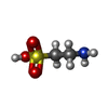 ChemComp-TAU:  ChemComp-PEG:  ChemComp-PG4:  ChemComp-ACT:  ChemComp-MLI:  ChemComp-EDO: |
| Source |
|
 Keywords Keywords | HYDROLASE / redox-regulation / glycosyl hydrolase / mannosidase / redox-switch / metagenome / disulfide bond |


