Cristina M Risi / Maicon Landim-Vieira / Betty Belknap / P Bryant Chase / Jose R Pinto / Vitold E Galkin /
PubMed Abstract
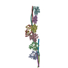
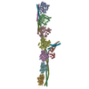
Cardiac muscle contraction/relaxation cycle depends on the rising and falling Ca levels in sarcomeres that control the extent of interactions between myosin-based thick and actin-based thin filaments. ...Cardiac muscle contraction/relaxation cycle depends on the rising and falling Ca levels in sarcomeres that control the extent of interactions between myosin-based thick and actin-based thin filaments. Cardiac thin filament (cTF) consists of actin, tropomyosin (Tm) that regulates myosin binding to actin, and troponin complex that governs Tm position upon Ca-binding. Troponin has three subunits - Ca-binding troponin C (TnC), Tm stabilizing troponin T (TnT), and inhibitory troponin I (TnI). TnT N-terminus (TnT1) interactions with actin stabilize the inhibited state of cTF. TnC, TnI, and Tm work in concert to control actomyosin interactions. Cryo-electron microscopy (cryo-EM) provided factual structures of healthy cTF, but structures of cTF carrying missense mutations linked to human cardiomyopathy are unknown. Variant Ile79Asn in human cardiac TnT (TnT-I79N) increases myofilament Ca sensitivity and slows cross-bridge kinetics, leading to severe hypertrophic/restrictive cardiomyopathy. Here, we used TnT-I79N mutation as a tool to examine the role of TnT1 in the complex mechanism of cTF regulation. Comparison of the cryo-EM structures of murine wild type and TnT-I79N native cTFs at systolic Ca levels (pCa = 5.8) demonstrates that TnT-I79N causes 1) dissociation of the TnT1 loop from its actin interface that results in Tm release to a more activated position, 2) reduced interaction of TnI C-terminus with actin-Tm, and 3) increased frequency of Ca-bound regulatory units. Our data indicate that the TnT1 loop is a crucial element of the allosteric regulatory network that couples Tn subunits and Tm to maintain adequate cTF response to physiological Ca levels during a heartbeat.
EMDB-47449, PDB-9e2e: The structure of the junction region of the wild-type murine native cardiac thin filament in Ca2+-free state Method: EM (single particle) / Resolution: 4.0 Å
EMDB-48447, PDB-9mo4: Structure of native murine cardiac thin filament at pCa=5.8 in Ca2+-free state (lower strand) Method: EM (single particle) / Resolution: 5.3 Å
EMDB-48448, PDB-9mo5: Structure of native murine cardiac thin filament at pCa=5.8 in Ca2+-bound partially activated state (lower strand) Method: EM (single particle) / Resolution: 5.3 Å
EMDB-48449, PDB-9mo6: Structure of native murine cardiac thin filament at pCa=5.8 in Ca2+-free tilted state (lower strand) Method: EM (single particle) / Resolution: 5.3 Å
EMDB-48450, PDB-9mo7: Structure of native murine cardiac thin filament at pCa=5.8 in Ca2+-bound fully activated state (lower strand) Method: EM (single particle) / Resolution: 5.2 Å
EMDB-48451, PDB-9mo8: Structure of native murine cardiac thin filament at pCa=5.8 in Ca2+-free state (upper strand) Method: EM (single particle) / Resolution: 5.2 Å
EMDB-48452, PDB-9mo9: Structure of native murine cardiac thin filament variant I79N in troponin T at pCa=5.8 in Ca2+-free rotated state (lower strand) Method: EM (single particle) / Resolution: 5.6 Å
EMDB-48453, PDB-9moa: Structure of native murine cardiac thin filament variant I79N in troponin T at pCa=5.8 in Ca2+-free tilted state (lower strand) Method: EM (single particle) / Resolution: 5.4 Å
EMDB-48454, PDB-9mob: Structure of native murine cardiac thin filament variant I79N in troponin T at pCa=5.8 in Ca2+-free state (upper strand) Method: EM (single particle) / Resolution: 5.5 Å
EMDB-48455, PDB-9moc: Structure of native murine cardiac thin filament variant I79N in troponin T at pCa=5.8 in Ca2+-free rotated state (upper strand) Method: EM (single particle) / Resolution: 5.6 Å
EMDB-48456, PDB-9mod: Structure of native murine cardiac thin filament variant I79N in troponin T at pCa=5.8 in Ca2+-free tilted state (upper strand) Method: EM (single particle) / Resolution: 5.7 Å
EMDB-48467, PDB-9moi: Structure of native murine cardiac thin filament at pCa=5.8 in Ca2+-free rotated state (lower strand) Method: EM (single particle) / Resolution: 5.4 Å
EMDB-48468, PDB-9mok: Structure of native murine cardiac thin filament at pCa=5.8 in Ca2+-free rotated state (upper strand) Method: EM (single particle) / Resolution: 5.6 Å
EMDB-48469, PDB-9mol: Structure of native murine cardiac thin filament at pCa=5.8 in Ca2+-free tilted state (upper strand) Method: EM (single particle) / Resolution: 5.2 Å
EMDB-48470, PDB-9mom: Structure of native murine cardiac thin filament at pCa=5.8 in Ca2+-bound partially activated state (upper strand) Method: EM (single particle) / Resolution: 5.1 Å
EMDB-48471, PDB-9mon: Structure of native murine cardiac thin filament at pCa=5.8 in Ca2+-bound fully activated state (upper strand) Method: EM (single particle) / Resolution: 5.2 Å
EMDB-48476, PDB-9moo: Structure of native murine cardiac thin filament variant I79N in troponin T at pCa=5.8 in Ca2+-free state (lower strand) Method: EM (single particle) / Resolution: 5.2 Å
EMDB-48477, PDB-9mop: Structure of native murine cardiac thin filament variant I79N in troponin T at pCa=5.8 in Ca2+-bound fully activated state (lower strand) Method: EM (single particle) / Resolution: 5.0 Å
EMDB-48482, PDB-9mou: Structure of native murine cardiac thin filament variant I79N in troponin T at pCa=5.8 in Ca2+-bound partially activated state (upper strand) Method: EM (single particle) / Resolution: 5.6 Å
EMDB-48483, PDB-9mow: Structure of native murine cardiac thin filament variant I79N in troponin T at pCa=5.8 in Ca2+-bound fully activated state (upper strand) Method: EM (single particle) / Resolution: 4.9 Å
EMDB-48484, PDB-9mox: Structure of native murine cardiac thin filament variant I79N in troponin T at pCa=5.8 in Ca2+-bound partially activated state (lower strand) Method: EM (single particle) / Resolution: 5.2 Å
In the structure databanks used in Yorodumi, some data are registered as the other names, "COVID-19 virus" and "2019-nCoV". Here are the details of the virus and the list of structure data.
Jan 31, 2019. EMDB accession codes are about to change! (news from PDBe EMDB page)
EMDB accession codes are about to change! (news from PDBe EMDB page)
The allocation of 4 digits for EMDB accession codes will soon come to an end. Whilst these codes will remain in use, new EMDB accession codes will include an additional digit and will expand incrementally as the available range of codes is exhausted. The current 4-digit format prefixed with “EMD-” (i.e. EMD-XXXX) will advance to a 5-digit format (i.e. EMD-XXXXX), and so on. It is currently estimated that the 4-digit codes will be depleted around Spring 2019, at which point the 5-digit format will come into force.
The EM Navigator/Yorodumi systems omit the EMD- prefix.
Related info.:Q: What is EMD? / ID/Accession-code notation in Yorodumi/EM Navigator
 Movie
Movie Controller
 Structure viewers
 About Yorodumi Papers


 Authors
 External links


 Keywords