 ムービー
ムービー コントローラー
 構造ビューア
 万見文献について


+検索条件
-Structure paper
| タイトル | CryoET of β-amyloid and tau within postmortem Alzheimer's disease brain. |
|---|---|
| ジャーナル・号・ページ | Nature, Year 2024 |
| 掲載日 | 2024年7月10日 |
 著者 著者 | Madeleine A G Gilbert / Nayab Fatima / Joshua Jenkins / Thomas J O'Sullivan / Andreas Schertel / Yehuda Halfon / Martin Wilkinson / Tjado H J Morrema / Mirjam Geibel / Randy J Read / Neil A Ranson / Sheena E Radford / Jeroen J M Hoozemans / René A W Frank /    |
| PubMed 要旨 | A defining pathological feature of most neurodegenerative diseases is the assembly of proteins into amyloid that form disease-specific structures. In Alzheimer's disease, this is characterized by the ...A defining pathological feature of most neurodegenerative diseases is the assembly of proteins into amyloid that form disease-specific structures. In Alzheimer's disease, this is characterized by the deposition of β-amyloid and tau with disease-specific conformations. The in situ structure of amyloid in the human brain is unknown. Here, using cryo-fluorescence microscopy-targeted cryo-sectioning, cryo-focused ion beam-scanning electron microscopy lift-out and cryo-electron tomography, we determined in-tissue architectures of β-amyloid and tau pathology in a postmortem Alzheimer's disease donor brain. β-amyloid plaques contained a mixture of fibrils, some of which were branched, and protofilaments, arranged in parallel arrays and lattice-like structures. Extracellular vesicles and cuboidal particles defined the non-amyloid constituents of β-amyloid plaques. By contrast, tau inclusions formed parallel clusters of unbranched filaments. Subtomogram averaging a cluster of 136 tau filaments in a single tomogram revealed the polypeptide backbone conformation and filament polarity orientation of paired helical filaments within tissue. Filaments within most clusters were similar to each other, but were different between clusters, showing amyloid heterogeneity that is spatially organized by subcellular location. The in situ structural approaches outlined here for human donor tissues have applications to a broad range of neurodegenerative diseases. |
 リンク リンク | Nature / PubMed:38987603 |
| 手法 | EM (らせん対称) / EM (サブトモグラム平均) |
| 解像度 | 3.0 - 33.0 Å |
| 構造データ | 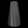 EMDB-18990: CryoEM map of tau PHF sarkosyl-extracted from a human AD patient (associated with in situ tomography) 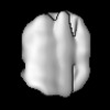 EMDB-50148: Tau PHF subtomogram average relating to CS1 extended data Figure 9A  EMDB-50152: Tau PHF subtomogram average relating to CS2 Figure 3i-j. 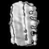 EMDB-50153: Tau PHF subtomogram average relating to CS3 extended data Figure 9c 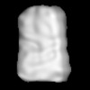 EMDB-50155: Tau PHF subtomogram average relating to CS4 extended data Figure 9d  EMDB-50156: Tau PHF subtomogram average relating to CS5 extended data Figure 9b  EMDB-50157: Tau PHF subtomogram average relating to CS6 extended data Figure 9e  EMDB-50159: Tau PHF subtomogram average relating to CS7 extended data Figure 9f 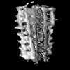 EMDB-50160: Tau PHF subtomogram average relating to LOL1_PHF Figure 4g-h  EMDB-50161: Tau SF subtomogram average relating to LOL1_SF Figure 4g-h 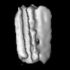 EMDB-50162: Tau SF subtomogram average relating to LOL2_SF Figure 4i-j |
| 由来 |
|
 Homo sapiens (ヒト)