ムービー
ムービー コントローラー
 構造ビューア
 万見文献について


+検索条件
-Structure paper
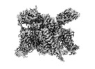
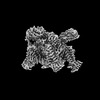
| タイトル | Unwinding and spiral sliding of S4 and domain rotation of VSD during the electromechanical coupling in Na1.7. |
|---|---|
| ジャーナル・号・ページ | Proc Natl Acad Sci U S A, Vol. 119, Issue 33, Page e2209164119, Year 2022 |
| 掲載日 | 2022年8月16日 |
 著者 著者 | Gaoxingyu Huang / Qiurong Wu / Zhangqiang Li / Xueqin Jin / Xiaoshuang Huang / Tong Wu / Xiaojing Pan / Nieng Yan /  |
| PubMed 要旨 | Voltage-gated sodium (Na) channel Na1.7 has been targeted for the development of nonaddictive pain killers. Structures of Na1.7 in distinct functional states will offer an advanced mechanistic ...Voltage-gated sodium (Na) channel Na1.7 has been targeted for the development of nonaddictive pain killers. Structures of Na1.7 in distinct functional states will offer an advanced mechanistic understanding and aid drug discovery. Here we report the cryoelectron microscopy analysis of a human Na1.7 variant that, with 11 rationally introduced point mutations, has a markedly right-shifted activation voltage curve with V reaching 69 mV. The voltage-sensing domain in the first repeat (VSD) in a 2.7-Å resolution structure displays a completely down (deactivated) conformation. Compared to the structure of WT Na1.7, three gating charge (GC) residues in VSD are transferred to the cytosolic side through a combination of helix unwinding and spiral sliding of S4 and ∼20° domain rotation. A conserved WNФФD motif on the cytoplasmic end of S3 stabilizes the down conformation of VSD. One GC residue is transferred in VSD mainly through helix sliding. Accompanying GC transfer in VSD and VSD, rearrangement and contraction of the intracellular gate is achieved through concerted movements of adjacent segments, including S4-5, S4-5, S5, and all S6 segments. Our studies provide important insight into the electromechanical coupling mechanism of the single-chain voltage-gated ion channels and afford molecular interpretations for a number of pain-associated mutations whose pathogenic mechanism cannot be revealed from previously reported Na structures. |
 リンク リンク | Proc Natl Acad Sci U S A / PubMed:35878056 / PubMed Central |
| 手法 | EM (単粒子) |
| 解像度 | 2.7 - 2.8 Å |
| 構造データ | EMDB-33484, PDB-7xve: EMDB-33485, PDB-7xvf: |
| 化合物 |  ChemComp-NAG:  ChemComp-Y01:  ChemComp-CLR:  ChemComp-LPE:  ChemComp-1PW:  ChemComp-PCW:  ChemComp-P5S: |
| 由来 |
|
 キーワード キーワード |  TRANSPORT PROTEIN (運搬体タンパク質) / Membrane protein. (膜タンパク質) / MEMBRANE PROTEIN (膜タンパク質) / action potential (活動電位) TRANSPORT PROTEIN (運搬体タンパク質) / Membrane protein. (膜タンパク質) / MEMBRANE PROTEIN (膜タンパク質) / action potential (活動電位) |