 Movie
Movie Controller
 Structure viewers
 About Yorodumi Papers


+Search query
-Structure paper
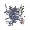
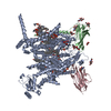
| Title | Unwinding and spiral sliding of S4 and domain rotation of VSD during the electromechanical coupling in Na1.7. |
|---|---|
| Journal, issue, pages | Proc Natl Acad Sci U S A, Vol. 119, Issue 33, Page e2209164119, Year 2022 |
| Publish date | Aug 16, 2022 |
 Authors Authors | Gaoxingyu Huang / Qiurong Wu / Zhangqiang Li / Xueqin Jin / Xiaoshuang Huang / Tong Wu / Xiaojing Pan / Nieng Yan /  |
| PubMed Abstract | Voltage-gated sodium (Na) channel Na1.7 has been targeted for the development of nonaddictive pain killers. Structures of Na1.7 in distinct functional states will offer an advanced mechanistic ...Voltage-gated sodium (Na) channel Na1.7 has been targeted for the development of nonaddictive pain killers. Structures of Na1.7 in distinct functional states will offer an advanced mechanistic understanding and aid drug discovery. Here we report the cryoelectron microscopy analysis of a human Na1.7 variant that, with 11 rationally introduced point mutations, has a markedly right-shifted activation voltage curve with V reaching 69 mV. The voltage-sensing domain in the first repeat (VSD) in a 2.7-Å resolution structure displays a completely down (deactivated) conformation. Compared to the structure of WT Na1.7, three gating charge (GC) residues in VSD are transferred to the cytosolic side through a combination of helix unwinding and spiral sliding of S4 and ∼20° domain rotation. A conserved WNФФD motif on the cytoplasmic end of S3 stabilizes the down conformation of VSD. One GC residue is transferred in VSD mainly through helix sliding. Accompanying GC transfer in VSD and VSD, rearrangement and contraction of the intracellular gate is achieved through concerted movements of adjacent segments, including S4-5, S4-5, S5, and all S6 segments. Our studies provide important insight into the electromechanical coupling mechanism of the single-chain voltage-gated ion channels and afford molecular interpretations for a number of pain-associated mutations whose pathogenic mechanism cannot be revealed from previously reported Na structures. |
 External links External links | Proc Natl Acad Sci U S A / PubMed:35878056 / PubMed Central |
| Methods | EM (single particle) |
| Resolution | 2.7 - 2.8 Å |
| Structure data | EMDB-33484, PDB-7xve: EMDB-33485, PDB-7xvf: |
| Chemicals |  ChemComp-NAG:  ChemComp-Y01:  ChemComp-CLR:  ChemComp-LPE:  ChemComp-1PW:  ChemComp-PCW:  ChemComp-P5S: |
| Source |
|
 Keywords Keywords | TRANSPORT PROTEIN / Membrane protein. / MEMBRANE PROTEIN / action potential |


 homo sapiens (human)