 Movie
Movie Controller
 Structure viewers
 About Yorodumi Papers


+Search query
-Structure paper
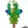
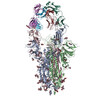
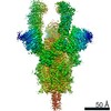
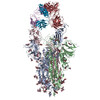
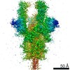
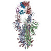
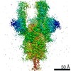
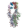
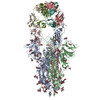
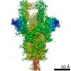
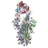
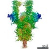
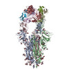
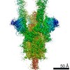
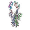
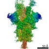
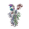
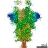
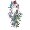
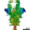
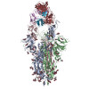
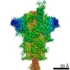
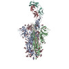
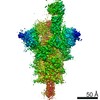
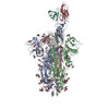
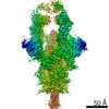
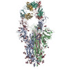
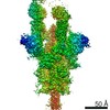
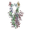
| Title | Structural basis for bivalent binding and inhibition of SARS-CoV-2 infection by human potent neutralizing antibodies. |
|---|---|
| Journal, issue, pages | Cell Res, Vol. 31, Issue 5, Page 517-525, Year 2021 |
| Publish date | Mar 17, 2021 |
 Authors Authors | Renhong Yan / Ruoke Wang / Bin Ju / Jinfang Yu / Yuanyuan Zhang / Nan Liu / Jia Wang / Qi Zhang / Peng Chen / Bing Zhou / Yaning Li / Yaping Shen / Shuyuan Zhang / Long Tian / Yingying Guo / Lu Xia / Xinyue Zhong / Lin Cheng / Xiangyang Ge / Juanjuan Zhao / Hong-Wei Wang / Xinquan Wang / Zheng Zhang / Linqi Zhang / Qiang Zhou /  |
| PubMed Abstract | Neutralizing monoclonal antibodies (nAbs) to severe acute respiratory syndrome coronavirus 2 (SARS-CoV-2) represent promising candidates for clinical intervention against coronavirus disease 2019 ...Neutralizing monoclonal antibodies (nAbs) to severe acute respiratory syndrome coronavirus 2 (SARS-CoV-2) represent promising candidates for clinical intervention against coronavirus disease 2019 (COVID-19). We isolated a large number of nAbs from SARS-CoV-2-infected individuals capable of disrupting proper interaction between the receptor binding domain (RBD) of the viral spike (S) protein and the receptor angiotensin converting enzyme 2 (ACE2). However, the structural basis for their potent neutralizing activity remains unclear. Here, we report cryo-EM structures of the ten most potent nAbs in their native full-length IgG-form or in both IgG-form and Fab-form bound to the trimeric S protein of SARS-CoV-2. The bivalent binding of the full-length IgG is found to associate with more RBDs in the "up" conformation than the monovalent binding of Fab, perhaps contributing to the enhanced neutralizing activity of IgG and triggering more shedding of the S1 subunit from the S protein. Comparison of a large number of nAbs identified common and unique structural features associated with their potent neutralizing activities. This work provides a structural basis for further understanding the mechanism of nAbs, especially through revealing the bivalent binding and its correlation with more potent neutralization and the shedding of S1 subunit. |
 External links External links | Cell Res / PubMed:33731853 / PubMed Central |
| Methods | EM (single particle) |
| Resolution | 2.7 - 7.3 Å |
| Structure data | EMDB-30512, PDB-7czp: EMDB-30513: cryo EM map of the S protein of SARS-CoV-2 in complex bound with P2B-1A10 EMDB-30514, PDB-7czr: EMDB-30515, PDB-7czs: EMDB-30516, PDB-7czt: EMDB-30517, PDB-7czu: EMDB-30518, PDB-7czv: EMDB-30519, PDB-7czw: EMDB-30520, PDB-7czx: EMDB-30521, PDB-7czy: EMDB-30522, PDB-7czz: EMDB-30523, PDB-7d00: EMDB-30524: S protein of SARS-CoV-2 in complex bound with P2B-1A10 EMDB-30529, PDB-7d0b: EMDB-30530, PDB-7d0c: EMDB-30531, PDB-7d0d: 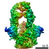 EMDB-30871: 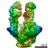 EMDB-30872: 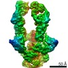 EMDB-30873: 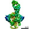 EMDB-30874: |
| Chemicals |  ChemComp-NAG: |
| Source |
|
 Keywords Keywords | VIRAL PROTEIN / SARS-CoV-2 / antibody / VIRAL PROTEIN/IMMUNE SYSTEM / Spike / IMMUNE SYSTEM / VIRAL PROTEIN-IMMUNE SYSTEM complex |


 homo sapiens (human)
 severe acute respiratory syndrome coronavirus 2