Movie
Movie Controller
 Structure viewers
 About Yorodumi Papers


+Search query
-Structure paper
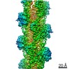
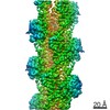
| Title | Cryo-electron microscopy structures of pyrene-labeled ADP-P- and ADP-actin filaments. |
|---|---|
| Journal, issue, pages | Nat Commun, Vol. 11, Issue 1, Page 5897, Year 2020 |
| Publish date | Nov 19, 2020 |
 Authors Authors | Steven Z Chou / Thomas D Pollard /  |
| PubMed Abstract | Since the fluorescent reagent N-(1-pyrene)iodoacetamide was first used to label skeletal muscle actin in 1981, the pyrene-labeled actin has become the most widely employed tool to measure the ...Since the fluorescent reagent N-(1-pyrene)iodoacetamide was first used to label skeletal muscle actin in 1981, the pyrene-labeled actin has become the most widely employed tool to measure the kinetics of actin polymerization and the interaction between actin and actin-binding proteins. Here we report high-resolution cryo-electron microscopy structures of actin filaments with N-1-pyrene conjugated to cysteine 374 and either ADP (3.2 Å) or ADP-phosphate (3.0 Å) in the active site. Polymerization buries pyrene in a hydrophobic cavity between subunits along the long-pitch helix with only minor differences in conformation compared with native actin filaments. These structures explain how polymerization increases the fluorescence 20-fold, how myosin and cofilin binding to filaments reduces the fluorescence, and how profilin binding to actin monomers increases the fluorescence. |
 External links External links | Nat Commun / PubMed:33214556 / PubMed Central |
| Methods | EM (helical sym.) |
| Resolution | 3.0 - 3.2 Å |
| Structure data | EMDB-22638, PDB-7k20: EMDB-22639, PDB-7k21: |
| Chemicals |  ChemComp-MG:  ChemComp-ADP:  ChemComp-1T4:  ChemComp-PO4: |
| Source |
|
 Keywords Keywords | CYTOSOLIC PROTEIN / actin / pyrene / fluorescence / ADP / phosphate |