 ムービー
ムービー コントローラー
 構造ビューア
 万見文献について


+検索条件
-Structure paper
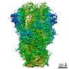
| タイトル | Cryo-EM structures of HKU2 and SADS-CoV spike glycoproteins provide insights into coronavirus evolution. |
|---|---|
| ジャーナル・号・ページ | Nat Commun, Vol. 11, Issue 1, Page 3070, Year 2020 |
| 掲載日 | 2020年6月17日 |
 著者 著者 | Jinfang Yu / Shuyuan Qiao / Runyu Guo / Xinquan Wang /  |
| PubMed 要旨 | Porcine coronavirus SADS-CoV has been identified from suckling piglets with severe diarrhea in southern China in 2017. The SADS-CoV genome shares ~95% identity to that of bat α-coronavirus HKU2, ...Porcine coronavirus SADS-CoV has been identified from suckling piglets with severe diarrhea in southern China in 2017. The SADS-CoV genome shares ~95% identity to that of bat α-coronavirus HKU2, suggesting that SADS-CoV may have emerged from a natural reservoir in bats. Here we report the cryo-EM structures of HKU2 and SADS-CoV spike (S) glycoprotein trimers at 2.38 Å and 2.83 Å resolution, respectively. We systematically compare the domains of HKU2 spike with those of α-, β-, γ-, and δ-coronavirus spikes, showing that the S1 subunit N- and C-terminal domains of HKU2/SADS-CoV are ancestral domains in the evolution of coronavirus spike proteins. The connecting region after the fusion peptide in the S2 subunit of HKU2/SADS-CoV adopts a unique conformation. These results structurally demonstrate a close evolutionary relationship between HKU2/SADS-CoV and β-coronavirus spikes and provide insights into the evolution and cross-species transmission of coronaviruses. |
 リンク リンク | Nat Commun / PubMed:32555182 / PubMed Central |
| 手法 | EM (単粒子) |
| 解像度 | 2.38 - 2.83 Å |
| 構造データ | EMDB-30037, PDB-6m15: EMDB-30038, PDB-6m16: |
| 化合物 |  ChemComp-NAG:  ChemComp-HOH: |
| 由来 |
|
 キーワード キーワード | VIRAL PROTEIN |


 rhinolophus bat coronavirus hku2 (ウイルス)