 Movie
Movie Controller
 Structure viewers
 About Yorodumi Papers


+Search query
-Structure paper
| Title | and phosphoribulokinase crystal structures complete the redox structural proteome of the Calvin-Benson cycle. |
|---|---|
| Journal, issue, pages | Proc Natl Acad Sci U S A, Vol. 116, Issue 16, Page 8048-8053, Year 2019 |
| Publish date | Apr 16, 2019 |
 Authors Authors | Libero Gurrieri / Alessandra Del Giudice / Nicola Demitri / Giuseppe Falini / Nicolae Viorel Pavel / Mirko Zaffagnini / Maurizio Polentarutti / Pierre Crozet / Christophe H Marchand / Julien Henri / Paolo Trost / Stéphane D Lemaire / Francesca Sparla / Simona Fermani /   |
| PubMed Abstract | In land plants and algae, the Calvin-Benson (CB) cycle takes place in the chloroplast, a specialized organelle in which photosynthesis occurs. Thioredoxins (TRXs) are small ubiquitous proteins, known ...In land plants and algae, the Calvin-Benson (CB) cycle takes place in the chloroplast, a specialized organelle in which photosynthesis occurs. Thioredoxins (TRXs) are small ubiquitous proteins, known to harmonize the two stages of photosynthesis through a thiol-based mechanism. Among the 11 enzymes of the CB cycle, the TRX target phosphoribulokinase (PRK) has yet to be characterized at the atomic scale. To accomplish this goal, we determined the crystal structures of PRK from two model species: the green alga (PRK) and the land plant (PRK). PRK is an elongated homodimer characterized by a large central β-sheet of 18 strands, extending between two catalytic sites positioned at its edges. The electrostatic surface potential of the catalytic cavity has both a positive region suitable for binding the phosphate groups of substrates and an exposed negative region to attract positively charged TRX-f. In the catalytic cavity, the regulatory cysteines are 13 Å apart and connected by a flexible region exclusive to photosynthetic eukaryotes-the clamp loop-which is believed to be essential for oxidation-induced structural rearrangements. Structural comparisons with prokaryotic and evolutionarily older PRKs revealed that both PRK and PRK have a strongly reduced dimer interface and an increased number of random-coiled regions, suggesting that a general loss in structural rigidity correlates with gains in TRX sensitivity during the molecular evolution of PRKs in eukaryotes. |
 External links External links | Proc Natl Acad Sci U S A / PubMed:30923119 / PubMed Central |
| Methods | SAS (X-ray synchrotron) / X-ray diffraction |
| Resolution | 2.471 - 2.6 Å |
| Structure data | 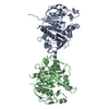 SASDDH9: 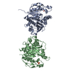 PDB-6h7g: 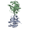 PDB-6h7h: |
| Chemicals |  ChemComp-SO4:  ChemComp-HOH: |
| Source |
|
 Keywords Keywords | PHOTOSYNTHESIS / Transferase / ATP binding / kinase activity / kinase |

 chlamydomonas reinhardtii (plant)