Dongsheng Lei / Yadong Yu / Yu-Lin Kuang / Jianfang Liu / Ronald M Krauss / Gang Ren /
PubMed Abstract
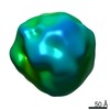
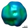
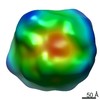
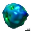
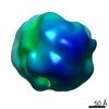
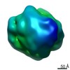
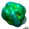
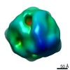
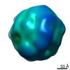
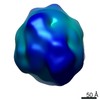
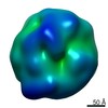
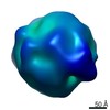
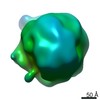
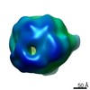
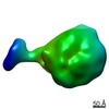
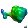
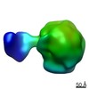
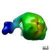
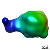
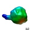
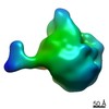
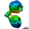
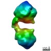
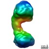
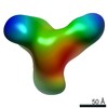
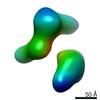
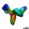
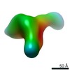
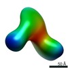
Intermediate-density lipoproteins (IDLs), the remnants of very-low-density lipoproteins via lipolysis, are rich in cholesteryl ester and are associated with cardiovascular disease. Despite ...Intermediate-density lipoproteins (IDLs), the remnants of very-low-density lipoproteins via lipolysis, are rich in cholesteryl ester and are associated with cardiovascular disease. Despite pharmacological interest in IDLs, their three-dimensional (3D) structure is still undetermined due to their variation in size, composition, and dynamic structure. To explore the 3D structure of IDLs, we reconstructed 3D density maps from individual IDL particles using cryo-electron microscopy (cryo-EM) and individual-particle electron tomography (IPET, without averaging from different molecules). 3D reconstructions of IDLs revealed an unexpected polyhedral structure that deviates from the generally assumed spherical shape model (Frias et al., 2007; Olson, 1998; Shen et al., 1977). The polyhedral-shaped IDL contains a high-density shell formed by flat surfaces that are similar to those of very-low-density lipoproteins but have sharper dihedral angles between nearby surfaces. These flat surfaces would be less hydrophobic than the curved surface of mature spherical high-density lipoprotein (HDL), leading to a lower binding affinity of IDL to hydrophobic proteins (such as cholesteryl ester transfer protein) than HDL. This is the first visualization of the IDL 3D structure, which could provide fundamental clues for delineating the role of IDL in lipid metabolism and cardiovascular disease.
EMDB-9069: Single-Molecule 3D Image of Human Plasma Intermediate-Density Lipoprotein (No. 01) Method: EM (tomography) / Resolution: 96.7 Å
EMDB-9070: Single-Molecule 3D Image of Human Plasma Intermediate-Density Lipoprotein (No. 02) Method: EM (tomography) / Resolution: 96.1 Å
EMDB-9071: Single-Molecule 3D Image of Human Plasma Intermediate-Density Lipoprotein (No. 03) Method: EM (tomography) / Resolution: 82.7 Å
EMDB-9072: Single-Molecule 3D Image of Human Plasma Intermediate-Density Lipoprotein (No. 04) Method: EM (tomography) / Resolution: 85.8 Å
EMDB-9073: Single-Molecule 3D Image of Human Plasma Intermediate-Density Lipoprotein (No. 05) Method: EM (tomography) / Resolution: 82.9 Å
EMDB-9074: Single-Molecule 3D Image of Human Plasma Intermediate-Density Lipoprotein (No. 06) Method: EM (tomography) / Resolution: 86.0 Å
EMDB-9075: Single-Molecule 3D Image of Human Plasma Intermediate-Density Lipoprotein (No. 07) Method: EM (tomography) / Resolution: 62.7 Å
EMDB-9076: Single-Molecule 3D Image of Human Plasma Intermediate-Density Lipoprotein (No. 08) Method: EM (tomography) / Resolution: 93.9 Å
EMDB-9077: Single-Molecule 3D Image of Human Plasma Intermediate-Density Lipoprotein (No. 09) Method: EM (tomography) / Resolution: 82.9 Å
EMDB-9078: Single-Molecule 3D Image of Human Plasma Intermediate-Density Lipoprotein (No. 10) Method: EM (tomography) / Resolution: 81.7 Å
EMDB-9079: Single-Molecule 3D Image of Human Plasma Intermediate-Density Lipoprotein (No. 11) Method: EM (tomography) / Resolution: 68.8 Å
EMDB-9080: Single-Molecule 3D Image of Human Plasma Intermediate-Density Lipoprotein (No. 12) Method: EM (tomography) / Resolution: 100.1 Å
EMDB-9081: Single-Molecule 3D Image of Human Plasma Intermediate-Density Lipoprotein (No. 13) Method: EM (tomography) / Resolution: 66.9 Å
EMDB-9082: Single-Molecule 3D Image of Human Plasma Intermediate-Density Lipoprotein (No. 14) Method: EM (tomography) / Resolution: 75.8 Å
EMDB-9083: Single-Molecule 3D Image of Human Plasma Intermediate-Density Lipoprotein (No. 15) Method: EM (tomography) / Resolution: 93.3 Å
EMDB-9084: Single-Molecule 3D Image of Human Plasma Intermediate-Density Lipoprotein (No. 16) Method: EM (tomography) / Resolution: 71.9 Å
EMDB-9085: Single-Molecule 3D Image of Human Plasma Intermediate-Density Lipoprotein in Complex with Monoclonal Antibody MAB012 (No. 01) Method: EM (tomography) / Resolution: 66.9 Å
EMDB-9086: Single-Molecule 3D Image of Human Plasma Intermediate-Density Lipoprotein in Complex with Monoclonal Antibody MAB012 (No. 02) Method: EM (tomography) / Resolution: 61.5 Å
EMDB-9087: Single-Molecule 3D Image of Human Plasma Intermediate-Density Lipoprotein in Complex with Monoclonal Antibody MAB012 (No. 03) Method: EM (tomography) / Resolution: 58.2 Å
EMDB-9088: Single-Molecule 3D Image of Human Plasma Intermediate-Density Lipoprotein in Complex with Monoclonal Antibody MAB012 (No. 04) Method: EM (tomography) / Resolution: 59.7 Å
EMDB-9089: Single-Molecule 3D Image of Human Plasma Intermediate-Density Lipoprotein in Complex with Monoclonal Antibody MAB012 (No. 05) Method: EM (tomography) / Resolution: 59.9 Å
EMDB-9090: Single-Molecule 3D Image of Human Plasma Intermediate-Density Lipoprotein in Complex with Monoclonal Antibody MAB012 (No. 06) Method: EM (tomography) / Resolution: 58.1 Å
EMDB-9091: Single-Molecule 3D Image of Human Plasma Intermediate-Density Lipoprotein in Complex with Monoclonal Antibody MAB012 (No. 07) Method: EM (tomography) / Resolution: 58.5 Å
EMDB-9092: Single-Molecule 3D Image of Two Human Plasma Intermediate-Density Lipoproteins in Complex with One Monoclonal Antibody MAB012 (No. 01) Method: EM (tomography) / Resolution: 60.6 Å
EMDB-9093: Single-Molecule 3D Image of Two Human Plasma Intermediate-Density Lipoproteins in Complex with One Monoclonal Antibody MAB012 (No. 02) Method: EM (tomography) / Resolution: 58.2 Å
EMDB-9094: Single-Molecule 3D Image of Two Human Plasma Intermediate-Density Lipoproteins in Complex with One Monoclonal Antibody MAB012 (No. 03) Method: EM (tomography) / Resolution: 68.6 Å
EMDB-9095: Single-Molecule 3D Image of Monoclonal Antibody MAB012 (No. 01) Method: EM (tomography) / Resolution: 59.9 Å
EMDB-9096: Single-Molecule 3D Image of Monoclonal Antibody MAB012 (No. 02) Method: EM (tomography) / Resolution: 56.5 Å
EMDB-9097: Single-Molecule 3D Image of Monoclonal Antibody MAB012 (No. 03) Method: EM (tomography) / Resolution: 58.4 Å
EMDB-9098: Single-Molecule 3D Image of Monoclonal Antibody MAB012 (No. 04) Method: EM (tomography) / Resolution: 61.0 Å
EMDB-9099: Single-Molecule 3D Image of Monoclonal Antibody MAB012 (No. 05) Method: EM (tomography) / Resolution: 58.1 Å
Source
Homo sapiens (human)
Mus musculus (house mouse)
+
About Yorodumi Papers
-
News
-
Feb 9, 2022. New format data for meta-information of EMDB entries
New format data for meta-information of EMDB entries
Version 3 of the EMDB header file is now the official format.
The previous official version 1.9 will be removed from the archive.
In the structure databanks used in Yorodumi, some data are registered as the other names, "COVID-19 virus" and "2019-nCoV". Here are the details of the virus and the list of structure data.
Jan 31, 2019. EMDB accession codes are about to change! (news from PDBe EMDB page)
EMDB accession codes are about to change! (news from PDBe EMDB page)
The allocation of 4 digits for EMDB accession codes will soon come to an end. Whilst these codes will remain in use, new EMDB accession codes will include an additional digit and will expand incrementally as the available range of codes is exhausted. The current 4-digit format prefixed with “EMD-” (i.e. EMD-XXXX) will advance to a 5-digit format (i.e. EMD-XXXXX), and so on. It is currently estimated that the 4-digit codes will be depleted around Spring 2019, at which point the 5-digit format will come into force.
The EM Navigator/Yorodumi systems omit the EMD- prefix.
Related info.:Q: What is EMD? / ID/Accession-code notation in Yorodumi/EM Navigator
 Movie
Movie Controller
 Structure viewers
 About Yorodumi Papers


 Authors
 External links
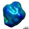

 Homo sapiens (human)
