 Movie
Movie Controller
 Structure viewers
 About Yorodumi Papers


+Search query
-Structure paper
| Title | Human MICAL1: Activation by the small GTPase Rab8 and small-angle X-ray scattering studies on the oligomerization state of MICAL1 and its complex with Rab8. |
|---|---|
| Journal, issue, pages | Protein Sci, Vol. 28, Issue 1, Page 150-166, Year 2019 |
| Publish date | Oct 31, 2018 |
 Authors Authors | Alessandro Esposito / Valeria Ventura / Maxim V Petoukhov / Amrita Rai / Dmitri I Svergun / Maria A Vanoni /    |
| PubMed Abstract | Human MICAL1 is a member of a recently discovered family of multidomain proteins that couple a FAD-containing monooxygenase-like domain to typical protein interaction domains. Growing evidence ...Human MICAL1 is a member of a recently discovered family of multidomain proteins that couple a FAD-containing monooxygenase-like domain to typical protein interaction domains. Growing evidence implicates the NADPH oxidase reaction catalyzed by the flavoprotein domain in generation of hydrogen peroxide as a second messenger in an increasing number of cell types and as a specific modulator of actin filaments stability. Several proteins of the Rab families of small GTPases are emerging as regulators of MICAL activity by binding to its C-terminal helical domain presumably shifting the equilibrium from the free - auto-inhibited - conformation to the active one. We here extend the characterization of the MICAL1-Rab8 interaction and show that indeed Rab8, in the active GTP-bound state, stabilizes the active MICAL1 conformation causing a specific four-fold increase of k of the NADPH oxidase reaction. Kinetic data and small-angle X-ray scattering (SAXS) measurements support the formation of a 1:1 complex between full-length MICAL1 and Rab8 with an apparent dissociation constant of approximately 8 μM. This finding supports the hypothesis that Rab8 is a physiological regulator of MICAL1 activity and shows how the protein region preceding the C-terminal Rab-binding domain may mask one of the Rab-binding sites detected with the isolated C-terminal fragment. SAXS-based modeling allowed us to propose the first model of the free full-length MICAL1, which is consistent with an auto-inhibited conformation in which the C-terminal region prevents catalysis by interfering with the conformational changes that are predicted to occur during the catalytic cycle. |
 External links External links | Protein Sci / PubMed:30242933 / PubMed Central |
| Methods | SAS (X-ray synchrotron) |
| Structure data |  SASDDR9:  SASDDS9: 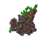 SASDDT9:  SASDDU9: |
| Source |
|
 Homo sapiens (human)