 Movie
Movie Controller
 Structure viewers
 About Yorodumi Papers


+Search query
-Structure paper
| Title | Optical and Structural Characterization of a Chronic Myeloid Leukemia DNA Biosensor. |
|---|---|
| Journal, issue, pages | ACS Chem Biol, Vol. 13, Issue 5, Page 1235-1242, Year 2018 |
| Publish date | May 18, 2018 |
 Authors Authors | Mílton Cordeiro / Ana Rita Otrelo-Cardoso / Dmitri I Svergun / Petr V Konarev / João Carlos Lima / Teresa Santos-Silva / Pedro Viana Baptista /    |
| PubMed Abstract | Selective base pairing is the foundation of DNA recognition. Here, we elucidate the molecular and structural details of a FRET-based two-component molecular beacon relying on steady-state ...Selective base pairing is the foundation of DNA recognition. Here, we elucidate the molecular and structural details of a FRET-based two-component molecular beacon relying on steady-state fluorescence spectroscopy, small-angle X-ray scattering (SAXS), microscale thermophoresis (MST), and differential electrophoretic mobility. This molecular beacon was designed to detect the most common fusion sequences causing chronic myeloid leukemia, e14a2 and e13a2. The emission spectra indicate that the self-assembly of the different components of the biosensor occurs sequentially, triggered by the fully complementary target. We further assessed the structural alterations leading to the specific fluorescence FRET signature by SAXS, MST, and the differential electrophoretic mobility, where the size range observed is consistent with hybridization and formation of a 1:1:1 complex for the probe in the presence of the complementary target and revelator. These results highlight the importance of different techniques to explore conformational DNA changes in solution and its potential to design and characterize molecular biosensors for genetic disease diagnosis. |
 External links External links | ACS Chem Biol / PubMed:29562136 |
| Methods | SAS (X-ray synchrotron) |
| Structure data |  SASDC95:  SASDCA5:  SASDCB5:  SASDCC5:  SASDCD5: 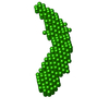 SASDCE5: 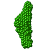 SASDCF5: 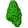 SASDCG5: 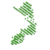 SASDCH5: 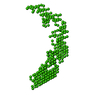 SASDCJ5: 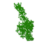 SASDCK5: 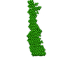 SASDCL5: |
