 Movie
Movie Controller
 Structure viewers
 About Yorodumi Papers


+Search query
-Structure paper
| Title | Electron microscopy and in vitro deneddylation reveal similar architectures and biochemistry of isolated human and Flag-mouse COP9 signalosome complexes. |
|---|---|
| Journal, issue, pages | Biochem Biophys Res Commun, Vol. 450, Issue 2, Page 991-997, Year 2014 |
| Publish date | Jul 25, 2014 |
 Authors Authors | Beate Rockel / Tilo Schmaler / Xiaohua Huang / Wolfgang Dubiel /  |
| PubMed Abstract | The COP9 signalosome (CSN) is a regulator of the ubiquitin (Ub) proteasome system (UPS). In the UPS, proteins are Ub-labeled for degradation by Ub ligases conferring substrate specificity. The CSN ...The COP9 signalosome (CSN) is a regulator of the ubiquitin (Ub) proteasome system (UPS). In the UPS, proteins are Ub-labeled for degradation by Ub ligases conferring substrate specificity. The CSN controls a large family of Ub ligases called cullin-RING ligases (CRLs), which ubiquitinate cell cycle regulators, transcription factors and DNA damage response proteins. The CSN possesses structural similarities with the 26S proteasome Lid complex and the translation initiation complex 3 (eIF3) indicating similar ancestry and function. Initial structures were obtained 14years ago by 2D electron microscopy (EM). Recently, first 3D molecular models of the CSN were created on the basis of negative-stain EM and single-particle analysis, mostly with recombinant complexes. Here, we compare deneddylating activity and structural features of CSN complexes purified in an elaborate procedure from human erythrocytes and efficiently pulled down from mouse Flag-CSN2 B8 fibroblasts. In an in vitro deneddylation assay both the human and the mouse CSN complexes deneddylated Nedd8-Cul1 with comparable rates. 3D structural models of the erythrocyte CSN as well as of the mouse Flag-CSN were generated by negative stain EM and by cryo-EM. Both complexes show a central U-shaped segment from which several arms emanate. This structure, called the horseshoe, is formed by the PCI domain subunits. CSN5 and CSN6 point away from the horseshoe. Compared to 3D models of negatively stained CSN complexes, densities assigned to CSN2 and CSN4 are better defined in the cryo-map. Because biochemical and structural results obtained with CSN complexes isolated from human erythrocytes and purified by Flag-CSN pulldown from mouse B8 fibroblasts are very similar, Flag-CSN pulldowns are a proper alternative to CSN preparation from erythrocytes. |
 External links External links | Biochem Biophys Res Commun / PubMed:24973710 |
| Methods | EM (single particle) |
| Resolution | 19.0 - 27.0 Å |
| Structure data |  EMDB-2569:  EMDB-2570: 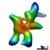 EMDB-2571: |
| Source |
|
 Homo sapiens (human)
