 Movie
Movie Controller
 Structure viewers
 About Yorodumi Papers


+Search query
-Structure paper
| Title | Solution structures of polcalcin Phl p 7 in three ligation states: Apo-, hemi-Mg2+-bound, and fully Ca2+-bound. |
|---|---|
| Journal, issue, pages | Proteins, Vol. 81, Issue 2, Page 300-315, Year 2013 |
| Publish date | Nov 12, 2012 |
 Authors Authors | Michael T Henzl / Arthur G Sirianni / Wei G Wycoff / Anmin Tan / John J Tanner /  |
| PubMed Abstract | Polcalcins are small EF-hand proteins believed to assist in regulating pollen-tube growth. Phl p 7, from timothy grass (Phleum pratense), crystallizes as a domain-swapped dimer at low pH. This study ...Polcalcins are small EF-hand proteins believed to assist in regulating pollen-tube growth. Phl p 7, from timothy grass (Phleum pratense), crystallizes as a domain-swapped dimer at low pH. This study describes the solution structures of the recombinant protein in buffered saline at pH 6.0, containing either 5.0 mM EDTA, 5.0 mM Mg(2+), or 100 μM Ca(2+). Phl p 7 is monomeric in all three ligation states. In the apo-form, both EF-hand motifs reside in the closed conformation, with roughly antiparallel N- and C-terminal helical segments. In 5.0 mM Mg(2+), the divalent ion is bound by EF-hand 2, perturbing interhelical angles and imposing more regular helical structure. The structure of Ca(2+)-bound Phl p 7 resembles that previously reported for Bet v 4-likewise exposing apolar surface to the solvent. Occluded in the apo- and Mg(2+)-bound forms, this surface presumably provides the docking site for Phl p 7 targets. Unlike Bet v 4, EF-hand 2 in Phl p 7 includes five potential anionic ligands, due to replacement of the consensus serine residue at -x (residue 55 in Phl p 7) with aspartate. In the Phl p 7 crystal structure, D55 functions as a helix cap for helix D. In solution, however, D55 apparently serves as a ligand to the bound Ca(2+). When Mg(2+) resides in site 2, the D55 carboxylate withdraws to a distance consistent with a role as an outer-sphere ligand. (15)N relaxation data, collected at 600 MHz, indicate that backbone mobility is limited in all three ligation states. |
 External links External links | Proteins / PubMed:23011803 |
| Methods | SAS (X-ray synchrotron) / NMR (solution) |
| Structure data |  SASDDJ2:  PDB-2lvi: 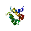 PDB-2lvj: 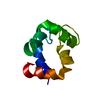 PDB-2lvk: |
| Chemicals |  ChemComp-MG:  ChemComp-CA: |
| Source |
|
 Keywords Keywords | ALLERGEN / EF-hand protein / calcium-binding protein / magnesium-binding protein |
 phleum pratense (timothy grass)