 Movie
Movie Controller
 Structure viewers
 About Yorodumi Papers


+Search query
-Structure paper
| Title | A molecular model of phosphorylation-based activation and potentiation of tarantula muscle thick filaments. |
|---|---|
| Journal, issue, pages | J Mol Biol, Vol. 414, Issue 1, Page 44-61, Year 2011 |
| Publish date | Nov 18, 2011 |
 Authors Authors | Reicy Brito / Lorenzo Alamo / Ulf Lundberg / José R Guerrero / Antonio Pinto / Guidenn Sulbarán / Mary Ann Gawinowicz / Roger Craig / Raúl Padrón /  |
| PubMed Abstract | Myosin filaments from many muscles are activated by phosphorylation of their regulatory light chains (RLCs). To elucidate the structural mechanism of activation, we have studied RLC phosphorylation ...Myosin filaments from many muscles are activated by phosphorylation of their regulatory light chains (RLCs). To elucidate the structural mechanism of activation, we have studied RLC phosphorylation in tarantula thick filaments, whose high-resolution structure is known. In the relaxed state, tarantula RLCs are ~50% non-phosphorylated and 50% mono-phosphorylated, while on activation, mono-phosphorylation increases, and some RLCs become bi-phosphorylated. Mass spectrometry shows that relaxed-state mono-phosphorylation occurs on Ser35, while Ca(2+)-activated phosphorylation is on Ser45, both located near the RLC N-terminus. The sequences around these serines suggest that they are the targets for protein kinase C and myosin light chain kinase (MLCK), respectively. The atomic model of the tarantula filament shows that the two myosin heads ("free" and "blocked") are in different environments, with only the free head serines readily accessible to kinases. Thus, protein kinase C Ser35 mono-phosphorylation in relaxed filaments would occur only on the free heads. Structural considerations suggest that these heads are less strongly bound to the filament backbone and may oscillate occasionally between attached and detached states ("swaying" heads). These heads would be available for immediate actin interaction upon Ca(2)(+) activation of the thin filaments. Once MLCK becomes activated, it phosphorylates free heads on Ser45. These heads become fully mobile, exposing blocked head Ser45 to MLCK. This would release the blocked heads, allowing their interaction with actin. On this model, twitch force would be produced by rapid interaction of swaying free heads with activated thin filaments, while prolonged exposure to Ca(2+) on tetanus would recruit new MLCK-activated heads, resulting in force potentiation. |
 External links External links | J Mol Biol / PubMed:21959262 / PubMed Central |
| Methods | EM (helical sym.) |
| Resolution | 20.0 Å |
| Structure data | 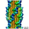 EMDB-1950: |
| Source |
|
 Aphonopelma sp. (spider)