 ムービー
ムービー コントローラー
 構造ビューア
 万見文献について


+検索条件
-Structure paper
| タイトル | Quaternary structure of the specific p53-DNA complex reveals the mechanism of p53 mutant dominance. |
|---|---|
| ジャーナル・号・ページ | Nucleic Acids Res, Vol. 39, Issue 20, Page 8960-8971, Year 2011 |
| 掲載日 | 2011年11月1日 |
 著者 著者 | Ricardo Aramayo / Michael B Sherman / Kathryne Brownless / Rudi Lurz / Andrei L Okorokov / Elena V Orlova /  |
| PubMed 要旨 | The p53 tumour suppressor is a transcriptional activator that controls cell fate in response to various stresses. p53 can initiate cell cycle arrest, senescence and/or apoptosis via transactivation ...The p53 tumour suppressor is a transcriptional activator that controls cell fate in response to various stresses. p53 can initiate cell cycle arrest, senescence and/or apoptosis via transactivation of p53 target genes, thus preventing cancer onset. Mutations that impair p53 usually occur in the core domain and negate the p53 sequence-specific DNA binding. Moreover, these mutations exhibit a dominant negative effect on the remaining wild-type p53. Here, we report the cryo electron microscopy structure of the full-length p53 tetramer bound to a DNA-encoding transcription factor response element (RE) at a resolution of 21 A. While two core domains from both dimers of the p53 tetramer interact with DNA within the complex, the other two core domains remain available for binding another DNA site. This finding helps to explain the dominant negative effect of p53 mutants based on the fact that p53 dimers are formed co-translationally before the whole tetramer assembles; therefore, a single mutant dimer would prevent the p53 tetramer from binding DNA. The structure indicates that the Achilles' heel of p53 is in its dimer-of-dimers organization, thus the tetramer activity can be negated by mutation in only one allele followed by tumourigenesis. |
 リンク リンク | Nucleic Acids Res / PubMed:21764777 / PubMed Central |
| 手法 | EM (単粒子) |
| 解像度 | 21.0 Å |
| 構造データ | 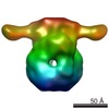 EMDB-1896: |
| 由来 |
|

