 Movie
Movie Controller
 Structure viewers
 About Yorodumi Papers


+Search query
-Structure paper
| Title | Comparison of Alzheimer Abeta(1-40) and Abeta(1-42) amyloid fibrils reveals similar protofilament structures. |
|---|---|
| Journal, issue, pages | Proc Natl Acad Sci U S A, Vol. 106, Issue 47, Page 19813-19818, Year 2009 |
| Publish date | Nov 24, 2009 |
 Authors Authors | Matthias Schmidt / Carsten Sachse / Walter Richter / Chen Xu / Marcus Fändrich / Nikolaus Grigorieff /  |
| PubMed Abstract | We performed mass-per-length (MPL) measurements and electron cryomicroscopy (cryo-EM) with 3D reconstruction on an Abeta(1-42) amyloid fibril morphology formed under physiological pH conditions. The ...We performed mass-per-length (MPL) measurements and electron cryomicroscopy (cryo-EM) with 3D reconstruction on an Abeta(1-42) amyloid fibril morphology formed under physiological pH conditions. The data show that the examined Abeta(1-42) fibril morphology has only one protofilament, although two protofilaments were observed with a previously studied Abeta(1-40) fibril. The latter fibril was resolved at 8 A resolution showing pairs of beta-sheets at the cores of the two protofilaments making up a fibril. Detailed comparison of the Abeta(1-42) and Abeta(1-40) fibril structures reveals that they share an axial twofold symmetry and a similar protofilament structure. Furthermore, the MPL data indicate that the protofilaments of the examined Abeta(1-40) and Abeta(1-42) fibrils have the same number of Abeta molecules per cross-beta repeat. Based on this data and the previously studied Abeta(1-40) fibril structure, we describe a model for the arrangement of peptides within the Abeta(1-42) fibril. |
 External links External links | Proc Natl Acad Sci U S A / PubMed:19843697 / PubMed Central |
| Methods | EM (helical sym.) |
| Resolution | 15.0 - 23.0 Å |
| Structure data |  EMDB-1649: 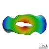 EMDB-1650: |
| Source |
|
 Homo sapiens (human)