+ Open data
- Basic information
| Entry | Database: EMDB / ID: EMD-9676 | |||||||||
|---|---|---|---|---|---|---|---|---|---|---|
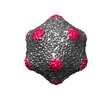
| Title | Vibrio phage M4 capsid | |||||||||
 Map data Map data | The icosahedral capsid of an El Tor vibriophage M4. | |||||||||
 Sample Sample |
| |||||||||
| Biological species |  Viruses Viruses | |||||||||
| Method | single particle reconstruction / cryo EM / Resolution: 15.4 Å | |||||||||
 Authors Authors | Das S / Dutta M / Sen A / Ghosh AN | |||||||||
| Funding support |  India, 1 items India, 1 items
| |||||||||
 Citation Citation |  Journal: Arch Virol / Year: 2019 Journal: Arch Virol / Year: 2019Title: Structural analysis and proteomics studies on the Myoviridae vibriophage M4. Authors: Sayani Das / Moumita Dutta / Anindito Sen / Amar N Ghosh /   Abstract: Bacteriophages play a crucial role in tracking the spread of bacterial epidemics. The frequent emergence of antibiotic-resistant bacterial strains throughout the world has motivated studies on ...Bacteriophages play a crucial role in tracking the spread of bacterial epidemics. The frequent emergence of antibiotic-resistant bacterial strains throughout the world has motivated studies on bacteriophages that can potentially be used in phage therapy as an alternative to conventional antibiotic treatment. A recent outbreak of cholera in Haiti took many lives due to a rapid development of resistance to the available antibiotics. The properties of vibriophages, bacteriophages that infect Vibrio cholerae, are therefore of practical interest. A detailed understanding of the structure and assembly of a vibriophage is potentially useful in developing phage therapy against cholera as well as for fabricating artificial nanocontainers. Therefore, the aim of the present study was to determine the three-dimensional organization of vibriophage M4 at sub-nanometer resolution by electron microscopy and single-particle analysis techniques to facilitate its use as a therapeutic agent. We found that M4 has a large capsid with T = 13 icosahedral symmetry and a long contractile tail. This double-stranded DNA phage also contains a head-to-tail connector protein complex that joins the capsid to the tail and a prominent baseplate at the end of the tail. This study also provides information regarding the proteome of this phage, which is proteins similar to that of other Myoviridae phages, and most of the encoded proteins are structural proteins that form the exquisite architecture of this bacteriophage. | |||||||||
| History |
|
- Structure visualization
| Movie |
 Movie viewer Movie viewer |
|---|---|
| Structure viewer | EM map:  SurfView SurfView Molmil Molmil Jmol/JSmol Jmol/JSmol |
| Supplemental images |
- Downloads & links
-EMDB archive
| Map data |  emd_9676.map.gz emd_9676.map.gz | 31.3 MB |  EMDB map data format EMDB map data format | |
|---|---|---|---|---|
| Header (meta data) |  emd-9676-v30.xml emd-9676-v30.xml emd-9676.xml emd-9676.xml | 7.4 KB 7.4 KB | Display Display |  EMDB header EMDB header |
| Images |  emd_9676.png emd_9676.png | 106.5 KB | ||
| Archive directory |  http://ftp.pdbj.org/pub/emdb/structures/EMD-9676 http://ftp.pdbj.org/pub/emdb/structures/EMD-9676 ftp://ftp.pdbj.org/pub/emdb/structures/EMD-9676 ftp://ftp.pdbj.org/pub/emdb/structures/EMD-9676 | HTTPS FTP |
-Validation report
| Summary document |  emd_9676_validation.pdf.gz emd_9676_validation.pdf.gz | 77.8 KB | Display |  EMDB validaton report EMDB validaton report |
|---|---|---|---|---|
| Full document |  emd_9676_full_validation.pdf.gz emd_9676_full_validation.pdf.gz | 76.9 KB | Display | |
| Data in XML |  emd_9676_validation.xml.gz emd_9676_validation.xml.gz | 495 B | Display | |
| Arichive directory |  https://ftp.pdbj.org/pub/emdb/validation_reports/EMD-9676 https://ftp.pdbj.org/pub/emdb/validation_reports/EMD-9676 ftp://ftp.pdbj.org/pub/emdb/validation_reports/EMD-9676 ftp://ftp.pdbj.org/pub/emdb/validation_reports/EMD-9676 | HTTPS FTP |
-Related structure data
- Links
Links
| EMDB pages |  EMDB (EBI/PDBe) / EMDB (EBI/PDBe) /  EMDataResource EMDataResource |
|---|
- Map
Map
| File |  Download / File: emd_9676.map.gz / Format: CCP4 / Size: 178 MB / Type: IMAGE STORED AS FLOATING POINT NUMBER (4 BYTES) Download / File: emd_9676.map.gz / Format: CCP4 / Size: 178 MB / Type: IMAGE STORED AS FLOATING POINT NUMBER (4 BYTES) | ||||||||||||||||||||||||||||||||||||||||||||||||||||||||||||||||||||
|---|---|---|---|---|---|---|---|---|---|---|---|---|---|---|---|---|---|---|---|---|---|---|---|---|---|---|---|---|---|---|---|---|---|---|---|---|---|---|---|---|---|---|---|---|---|---|---|---|---|---|---|---|---|---|---|---|---|---|---|---|---|---|---|---|---|---|---|---|---|
| Annotation | The icosahedral capsid of an El Tor vibriophage M4. | ||||||||||||||||||||||||||||||||||||||||||||||||||||||||||||||||||||
| Projections & slices | Image control
Images are generated by Spider. | ||||||||||||||||||||||||||||||||||||||||||||||||||||||||||||||||||||
| Voxel size | X=Y=Z: 2.5918 Å | ||||||||||||||||||||||||||||||||||||||||||||||||||||||||||||||||||||
| Density |
| ||||||||||||||||||||||||||||||||||||||||||||||||||||||||||||||||||||
| Symmetry | Space group: 1 | ||||||||||||||||||||||||||||||||||||||||||||||||||||||||||||||||||||
| Details | EMDB XML:
CCP4 map header:
| ||||||||||||||||||||||||||||||||||||||||||||||||||||||||||||||||||||
-Supplemental data
- Sample components
-Entire : Viruses
| Entire | Name:  Viruses Viruses |
|---|---|
| Components |
|
-Supramolecule #1: Viruses
| Supramolecule | Name: Viruses / type: virus / ID: 1 / Parent: 0 / Details: Vibrio phage M4, ATCC number-51354-B4 / NCBI-ID: 10239 / Sci species name: Viruses / Sci species strain: M4 / Virus type: VIRION / Virus isolate: OTHER / Virus enveloped: No / Virus empty: No |
|---|---|
| Host (natural) | Organism:  Vibrio cholerae MAK 757 (bacteria) Vibrio cholerae MAK 757 (bacteria) |
| Virus shell | Shell ID: 1 / Name: Vibrio phage M4 capsid / Diameter: 800.0 Å / T number (triangulation number): 13 |
-Experimental details
-Structure determination
| Method | cryo EM |
|---|---|
 Processing Processing | single particle reconstruction |
| Aggregation state | particle |
- Sample preparation
| Buffer | pH: 7.4 |
|---|---|
| Vitrification | Cryogen name: ETHANE / Instrument: LEICA EM CPC |
- Electron microscopy
| Microscope | FEI TECNAI 12 |
|---|---|
| Image recording | Film or detector model: KODAK SO-163 FILM / Average electron dose: 10.0 e/Å2 |
| Electron beam | Acceleration voltage: 120 kV / Electron source: TUNGSTEN HAIRPIN |
| Electron optics | Illumination mode: FLOOD BEAM / Imaging mode: BRIGHT FIELD |
| Sample stage | Cooling holder cryogen: NITROGEN |
- Image processing
| Final reconstruction | Resolution.type: BY AUTHOR / Resolution: 15.4 Å / Resolution method: FSC 0.143 CUT-OFF / Number images used: 500 |
|---|---|
| Initial angle assignment | Type: COMMON LINE |
| Final angle assignment | Type: COMMON LINE |
 Movie
Movie Controller


 UCSF Chimera


 Z (Sec.)
 Y (Row.)
 X (Col.)


