National Institutes of Health/National Institute Of Allergy and Infectious Diseases (NIH/NIAID)
R01AI089803
United States
National Institutes of Health/National Institute of General Medical Sciences (NIH/NIGMS)
T32 GM08061
United States
National Institutes of Health/National Institute Of Allergy and Infectious Diseases (NIH/NIAID)
R01 AIO56346
United States
National Institutes of Health/Office of the Director
S10 OD019995
United States
Citation
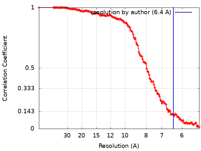
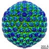
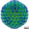
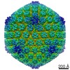
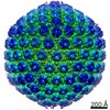
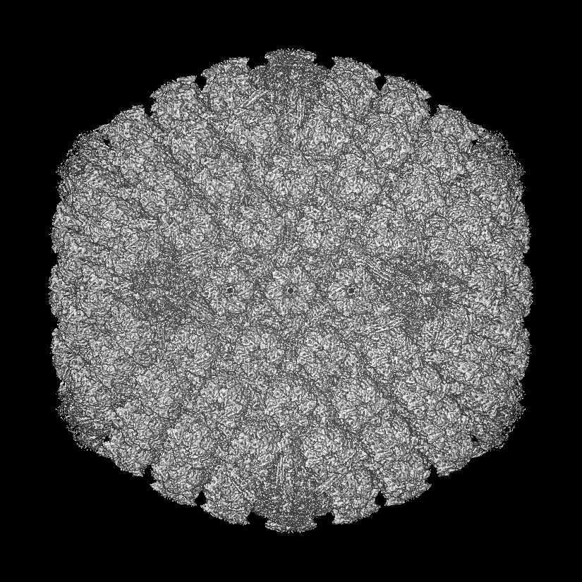
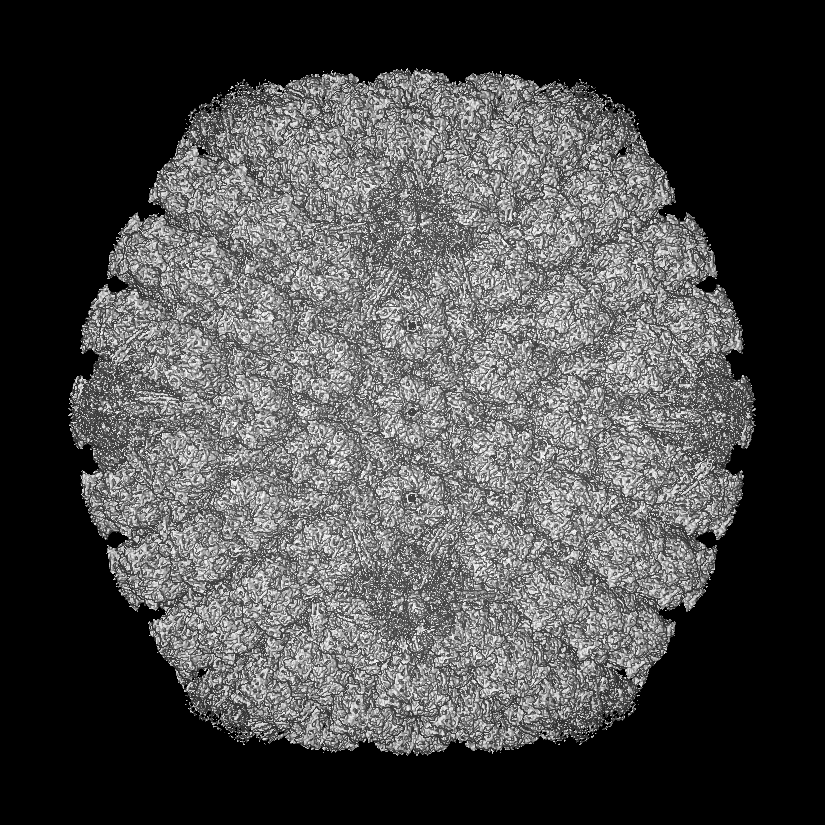
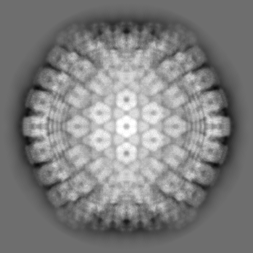
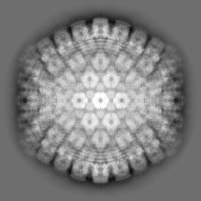
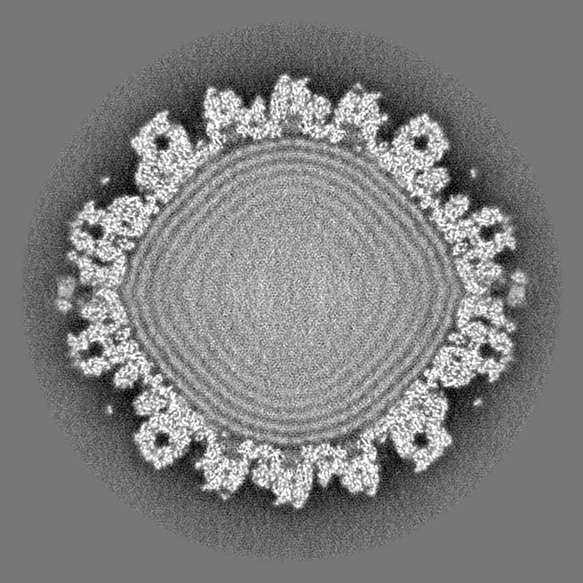
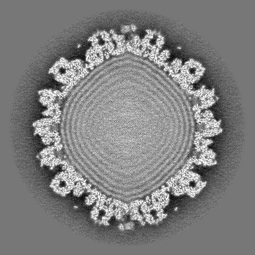
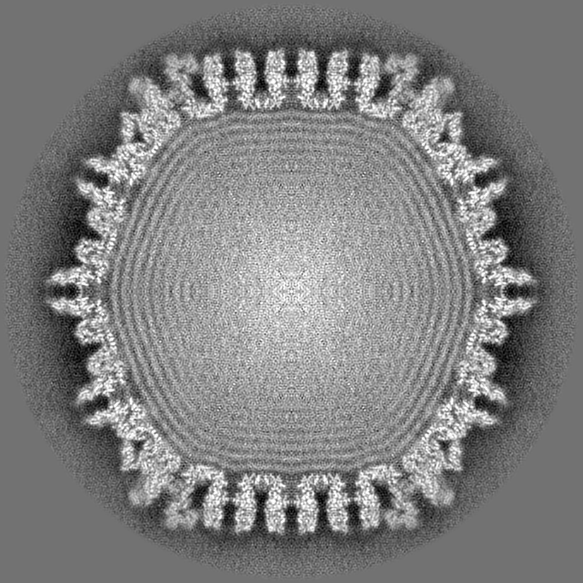
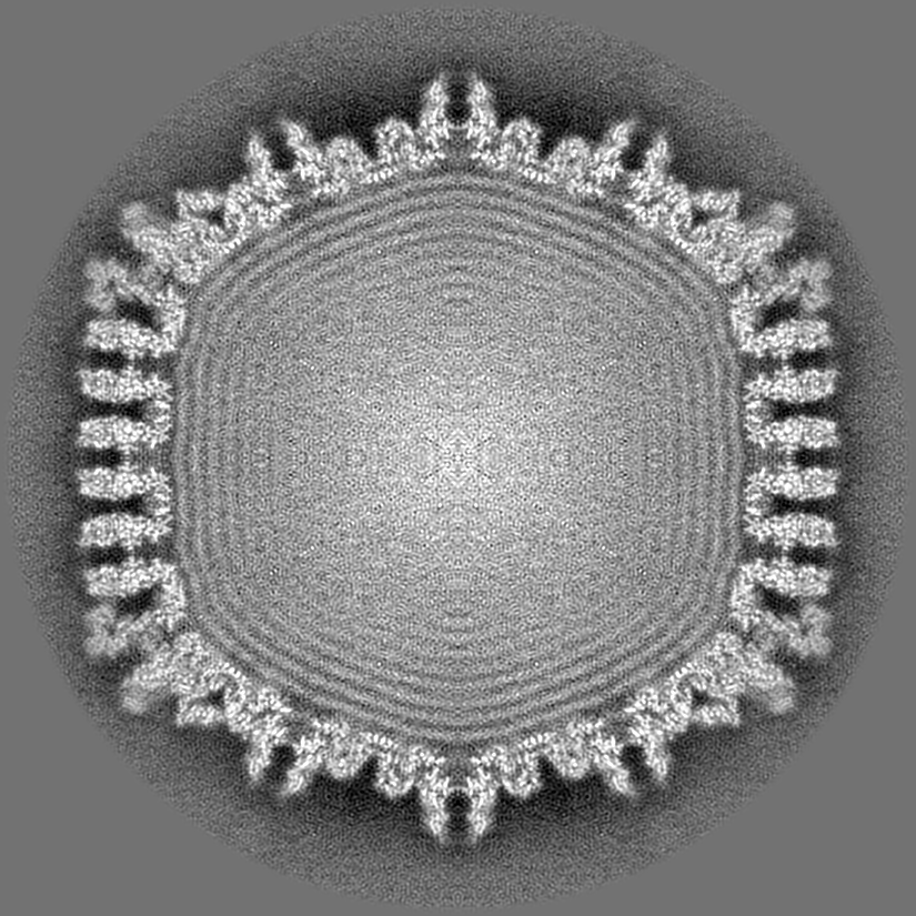
Journal: J Virol / Year: 2017 Title: The C Terminus of the Herpes Simplex Virus UL25 Protein Is Required for Release of Viral Genomes from Capsids Bound to Nuclear Pores. Authors: Jamie B Huffman / Gina R Daniel / Erik Falck-Pedersen / Alexis Huet / Greg A Smith / James F Conway / Fred L Homa / Abstract: The herpes simplex virus (HSV) capsid is released into the cytoplasm after fusion of viral and host membranes, whereupon dynein-dependent trafficking along microtubules targets it to the nuclear ...The herpes simplex virus (HSV) capsid is released into the cytoplasm after fusion of viral and host membranes, whereupon dynein-dependent trafficking along microtubules targets it to the nuclear envelope. Binding of the capsid to the nuclear pore complex (NPC) is mediated by the capsid protein pUL25 and the capsid-tethered tegument protein pUL36. Temperature-sensitive mutants in both pUL25 and pUL36 dock at the NPC but fail to release DNA. The uncoating reaction has been difficult to study due to the rapid release of the genome once the capsid interacts with the nuclear pore. In this study, we describe the isolation and characterization of a truncation mutant of pUL25. Live-cell imaging and immunofluorescence studies demonstrated that the mutant was not impaired in penetration of the host cell or in trafficking of the capsid to the nuclear membrane. However, expression of viral proteins was absent or significantly delayed in cells infected with the pUL25 mutant virus. Transmission electron microscopy revealed capsids accumulated at nuclear pores that retained the viral genome for at least 4 h postinfection. In addition, cryoelectron microscopy (cryo-EM) reconstructions of virion capsids did not detect any obvious differences in the location or structural organization for the pUL25 or pUL36 proteins on the pUL25 mutant capsids. Further, in contrast to wild-type virus, the antiviral response mediated by the viral DNA-sensing cyclic guanine adenine synthase (cGAS) was severely compromised for the pUL25 mutant. These results demonstrate that the pUL25 capsid protein has a critical role in releasing viral DNA from NPC-bound capsids. Herpes simplex virus 1 (HSV-1) is the causative agent of several pathologies ranging in severity from the common cold sore to life-threatening encephalitic infection. Early steps in infection include release of the capsid into the cytoplasm, docking of the capsid at a nuclear pore, and release of the viral genome into the nucleus. A key knowledge gap is how the capsid engages the NPC and what triggers release of the viral genome into the nucleus. Here we show that the C-terminal region of the HSV-1 pUL25 protein is required for releasing the viral genome from capsids docked at nuclear pores. The significance of our research is in identifying pUL25 as a key viral factor for genome uncoating. pUL25 is found at each of the capsid vertices as part of the capsid vertex-specific component and implicates the importance of this complex for NPC binding and genome release.
History
Deposition
May 15, 2017
-
Header (metadata) release
Jun 21, 2017
-
Map release
Jun 21, 2017
-
Update
Jan 29, 2020
-
Current status
Jan 29, 2020
Processing site: RCSB / Status: Released
-
Structure visualization
Movie
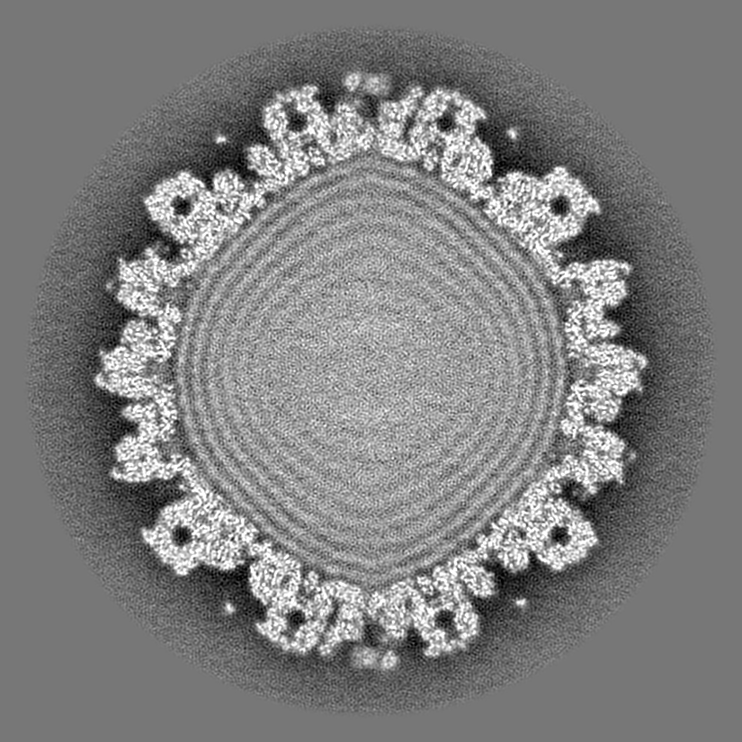
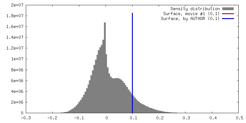
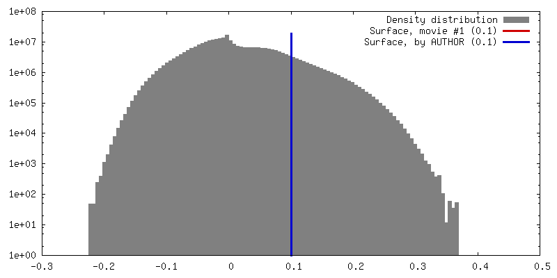
Surface view with section colored by density value
Virus: Human alphaherpesvirus 1 (Herpes simplex virus type 1)
-
Supramolecule #1: Human alphaherpesvirus 1
Supramolecule
Name: Human alphaherpesvirus 1 / type: virus / ID: 1 / Parent: 0 / NCBI-ID: 10298 / Sci species name: Human alphaherpesvirus 1 / Virus type: VIRION / Virus isolate: OTHER / Virus enveloped: Yes / Virus empty: No
Host (natural)
Organism: Homo sapiens (human)
Molecular weight
Theoretical: 144 MDa
Virus shell
Shell ID: 1 / Name: capsid / Diameter: 1200.0 Å / T number (triangulation number): 16
-
Experimental details
-
Structure determination
Method
cryo EM
Processing
single particle reconstruction
Aggregation state
particle
-
Sample preparation
Concentration
1 mg/mL
Buffer
pH: 7.5 Component:
Concentration
Name
Formula
10.0 mM
TRIS
150.0 mM
sodium chloride
NaCl
1.0 mM
EDTA
Grid
Model: Quantifoil R2/1 / Material: COPPER / Mesh: 400 / Support film - Material: CARBON / Support film - topology: CONTINUOUS / Pretreatment - Type: GLOW DISCHARGE
Vitrification
Cryogen name: ETHANE-PROPANE / Chamber humidity: 100 % / Chamber temperature: 293 K / Instrument: FEI VITROBOT MARK III / Details: 8 second blot.
Details
Virion
-
Electron microscopy
Microscope
FEI POLARA 300
Image recording
Film or detector model: FEI FALCON II (4k x 4k) / Digitization - Sampling interval: 14.0 µm / Digitization - Frames/image: 2-6 / Number grids imaged: 1 / Number real images: 4503 / Average exposure time: 1.3 sec. / Average electron dose: 10.0 e/Å2
Electron beam
Acceleration voltage: 300 kV / Electron source: FIELD EMISSION GUN
In the structure databanks used in Yorodumi, some data are registered as the other names, "COVID-19 virus" and "2019-nCoV". Here are the details of the virus and the list of structure data.
Jan 31, 2019. EMDB accession codes are about to change! (news from PDBe EMDB page)
EMDB accession codes are about to change! (news from PDBe EMDB page)
The allocation of 4 digits for EMDB accession codes will soon come to an end. Whilst these codes will remain in use, new EMDB accession codes will include an additional digit and will expand incrementally as the available range of codes is exhausted. The current 4-digit format prefixed with “EMD-” (i.e. EMD-XXXX) will advance to a 5-digit format (i.e. EMD-XXXXX), and so on. It is currently estimated that the 4-digit codes will be depleted around Spring 2019, at which point the 5-digit format will come into force.
The EM Navigator/Yorodumi systems omit the EMD- prefix.
Related info.:Q: What is EMD? / ID/Accession-code notation in Yorodumi/EM Navigator
Yorodumi is a browser for structure data from EMDB, PDB, SASBDB, etc.
This page is also the successor to EM Navigator detail page, and also detail information page/front-end page for Omokage search.
The word "yorodu" (or yorozu) is an old Japanese word meaning "ten thousand". "mi" (miru) is to see.
Related info.:EMDB / PDB / SASBDB / Comparison of 3 databanks / Yorodumi Search / Aug 31, 2016. New EM Navigator & Yorodumi / Yorodumi Papers / Jmol/JSmol / Function and homology information / Changes in new EM Navigator and Yorodumi
 Movie
Movie Controller

 Yorodumi
 Open data
 Basic information
 Map data
 Sample
 Human alphaherpesvirus 1 (Herpes simplex virus type 1)
 Authors
 United States, 4 items
  Citation
 Structure visualization
 Movie viewer
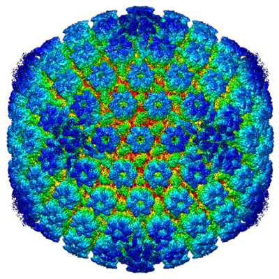
 Downloads & links
 emd_8733.png
 http://ftp.pdbj.org/pub/emdb/structures/EMD-8733

 Z (Sec.)
 Y (Row.)
 X (Col.)


 Sample components
 Homo sapiens (human)
 Processing
 Electron microscopy
 FIELD EMISSION GUN