 Movie
Movie Controller

[English] 日本語
 Yorodumi
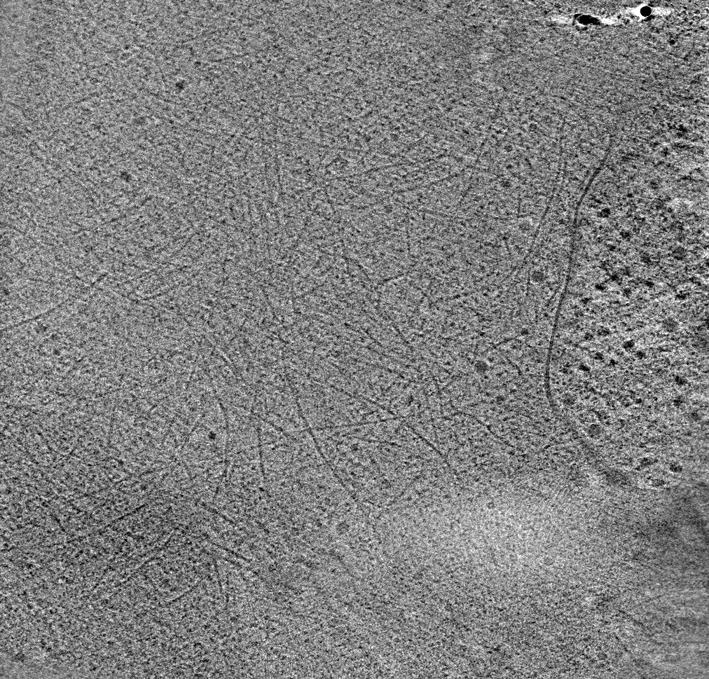
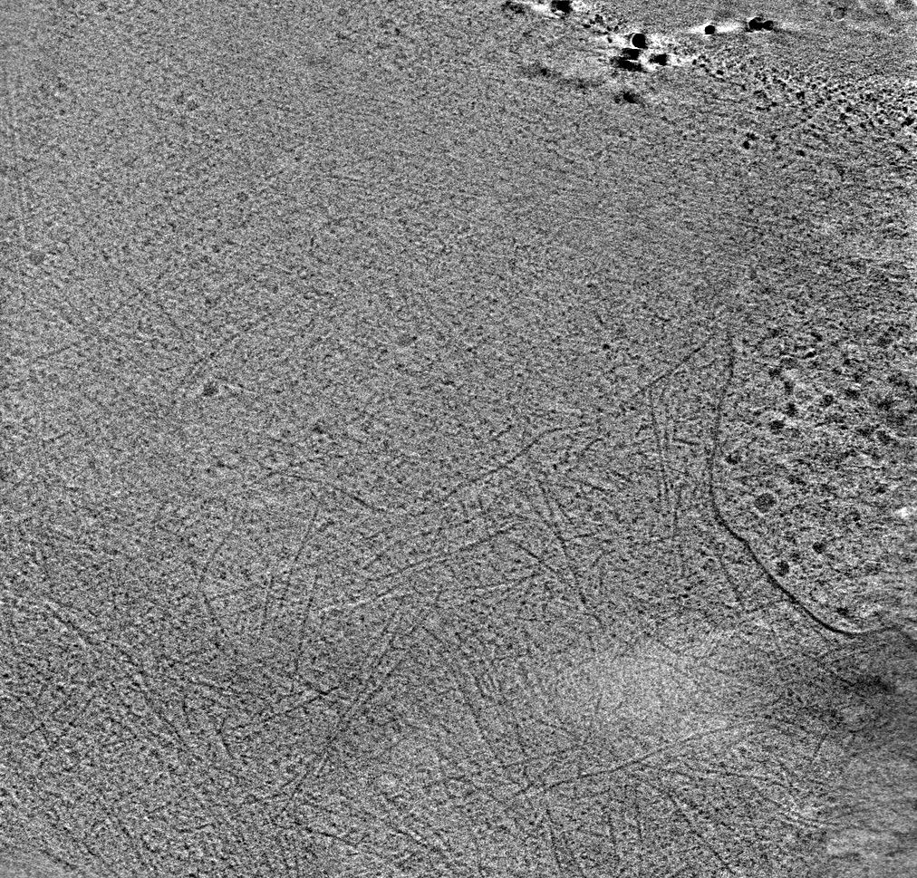
- EMDB-61092: Cryo-electron tomography of optogenetically-induced lamellipodia -
+ Open data

- Basic information
| Entry |  | ||||||||||||||||||||||||||||||||||||||||||
|---|---|---|---|---|---|---|---|---|---|---|---|---|---|---|---|---|---|---|---|---|---|---|---|---|---|---|---|---|---|---|---|---|---|---|---|---|---|---|---|---|---|---|---|
| Title | Cryo-electron tomography of optogenetically-induced lamellipodia | ||||||||||||||||||||||||||||||||||||||||||
 Map data Map data | |||||||||||||||||||||||||||||||||||||||||||
 Sample Sample |
| ||||||||||||||||||||||||||||||||||||||||||
 Keywords Keywords | actin cytoskeleton / plasma membrane / lamellipodia / STRUCTURAL PROTEIN | ||||||||||||||||||||||||||||||||||||||||||
| Biological species |  Chlorocebus aethiops (grivet) Chlorocebus aethiops (grivet) | ||||||||||||||||||||||||||||||||||||||||||
| Method | electron tomography / cryo EM | ||||||||||||||||||||||||||||||||||||||||||
 Authors Authors | Inaba H / Imasaki T / Aoyama K / Yoshihara S / Takazaki H / Kato T / Goto H / Mitsuoka K / Nitta R / Nakata T | ||||||||||||||||||||||||||||||||||||||||||
| Funding support |  Japan, 13 items Japan, 13 items
| ||||||||||||||||||||||||||||||||||||||||||
 Citation Citation | Journal: Biorxiv / Year: 2024 Title: Cryo-electron tomography of the actin cytoskeleton and membrane organization during lamellipodia formation using optogenetics. Authors: Inaba H / Imasaki T / Aoyama K / Yoshihara S / Takazaki H / Kato T / Goto H / Mitsuoka K / Nitta R / Nakata T | ||||||||||||||||||||||||||||||||||||||||||
| History |
|
- Structure visualization
| Supplemental images |
|---|

- Downloads & links
-EMDB archive
| Map data | emd_61092.map.gz | 527.1 MB |  EMDB map data format EMDB map data format | |
|---|---|---|---|---|
| Header (meta data) | emd-61092-v30.xmlemd-61092.xml | 11.4 KB 11.4 KB | Display Display | EMDB header |
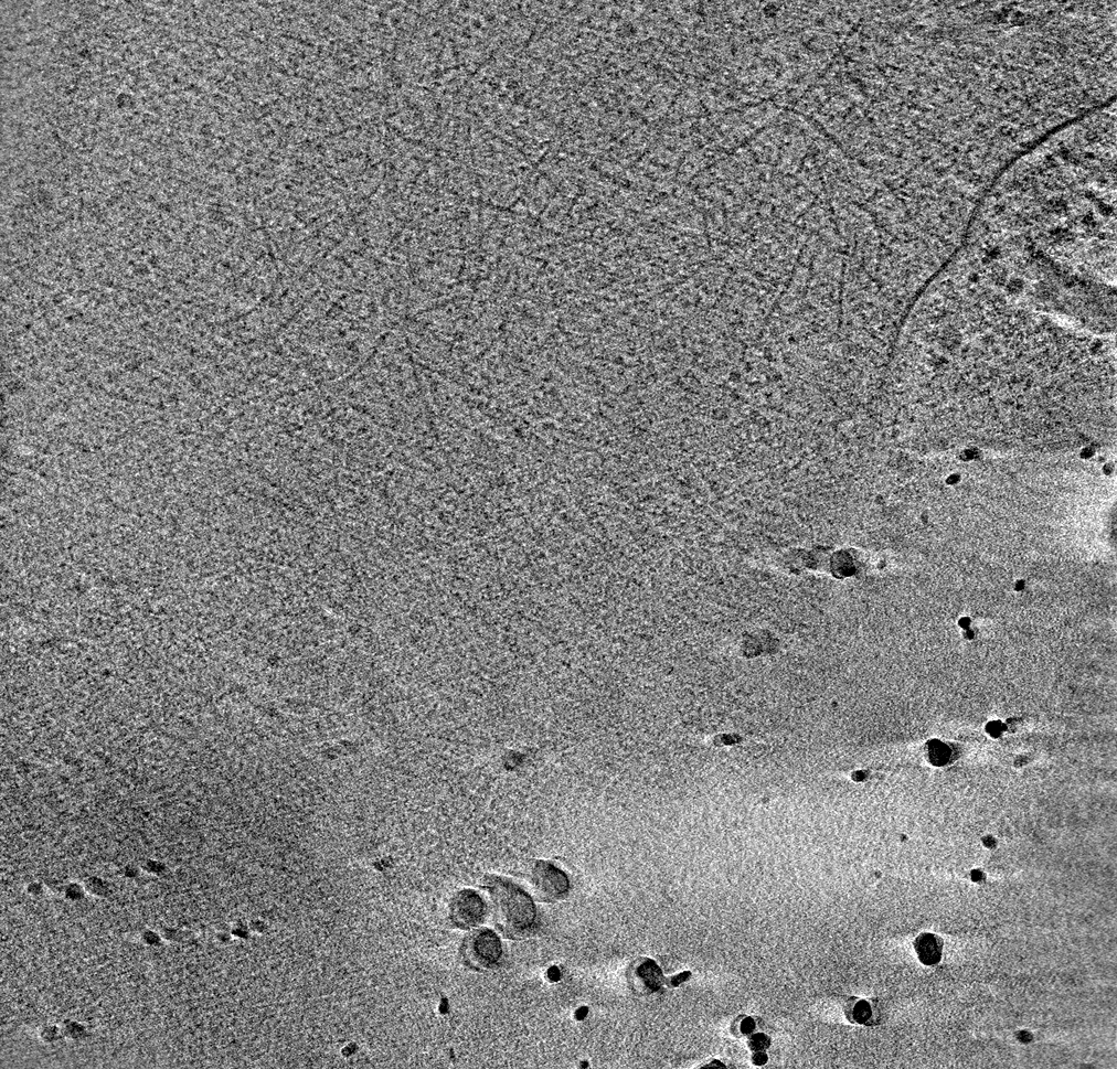
| Images |  emd_61092.png emd_61092.png | 209.4 KB | ||
| Filedesc metadata | emd-61092.cif.gz | 3.8 KB | ||
| Archive directory |  http://ftp.pdbj.org/pub/emdb/structures/EMD-61092ftp://ftp.pdbj.org/pub/emdb/structures/EMD-61092 http://ftp.pdbj.org/pub/emdb/structures/EMD-61092ftp://ftp.pdbj.org/pub/emdb/structures/EMD-61092 | HTTPS FTP |
-Validation report
| Summary document | emd_61092_validation.pdf.gz | 411.1 KB | Display | EMDB validaton report |
|---|---|---|---|---|
| Full document | emd_61092_full_validation.pdf.gz | 410.6 KB | Display | |
| Data in XML | emd_61092_validation.xml.gz | 5.2 KB | Display | |
| Data in CIF | emd_61092_validation.cif.gz | 5.8 KB | Display | |
| Arichive directory | https://ftp.pdbj.org/pub/emdb/validation_reports/EMD-61092ftp://ftp.pdbj.org/pub/emdb/validation_reports/EMD-61092 | HTTPS FTP |
-Related structure data

-Links
| EMDB pages | EMDB (EBI/PDBe) / EMDataResource |
|---|
-Map
| File | Download / File: emd_61092.map.gz / Format: CCP4 / Size: 568.6 MB / Type: IMAGE STORED AS FLOATING POINT NUMBER (4 BYTES) | ||||||||||||||||||||||||||||||||
|---|---|---|---|---|---|---|---|---|---|---|---|---|---|---|---|---|---|---|---|---|---|---|---|---|---|---|---|---|---|---|---|---|---|
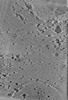
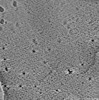
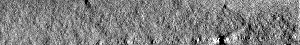
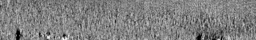
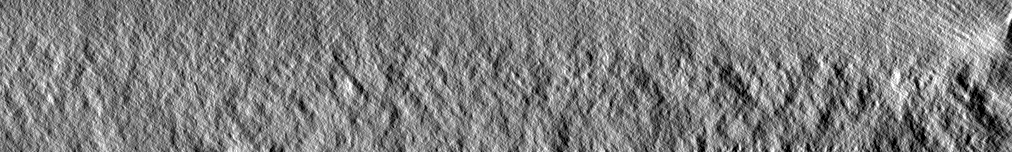
| Projections & slices | Image control
Images are generated by Spider. generated in cubic-lattice coordinate | ||||||||||||||||||||||||||||||||
| Voxel size | X=Y=Z: 14.8484 Å | ||||||||||||||||||||||||||||||||
| Density |
| ||||||||||||||||||||||||||||||||
| Symmetry | Space group: 1 | ||||||||||||||||||||||||||||||||
| Details | EMDB XML:
|
 Z (Sec.)
 Y (Row.)
 X (Col.)


-Supplemental data
- Sample components
-Entire : lamellipodia in COS-7 cells
| Entire | Name: lamellipodia in COS-7 cells |
|---|---|
| Components |
|
-Supramolecule #1: lamellipodia in COS-7 cells
| Supramolecule | Name: lamellipodia in COS-7 cells / type: cell / ID: 1 / Parent: 0 / Details: PA-Rac1 induced lamellipodia |
|---|---|
| Source (natural) | Organism: Chlorocebus aethiops (grivet) / Organ: kidney |
-Experimental details
-Structure determination
| Method | cryo EM |
|---|---|
 Processing Processing | electron tomography |
| Aggregation state | cell |
-Sample preparation
| Buffer | pH: 7.4 |
|---|---|
| Vitrification | Cryogen name: ETHANE |
| Sectioning | Other: NO SECTIONING |
| Fiducial marker | Manufacturer: SIGMA-Aldrich / Diameter: 20 nm |
- Electron microscopy
| Microscope | FEI TITAN KRIOS |
|---|---|
| Specialist optics | Phase plate: VOLTA PHASE PLATE |
| Image recording | Film or detector model: GATAN K3 BIOQUANTUM (6k x 4k) / Average electron dose: 1.44 e/Å2 |
| Electron beam | Acceleration voltage: 300 kV / Electron source:  FIELD EMISSION GUN FIELD EMISSION GUN |
| Electron optics | Illumination mode: FLOOD BEAM / Imaging mode: BRIGHT FIELD / Nominal defocus max: 4.0 µm / Nominal defocus min: 4.0 µm |
| Experimental equipment |  Model: Titan Krios / Image courtesy: FEI Company |
-Image processing
| Final reconstruction | Software - Name: IMOD (ver. 4.11.24) / Number images used: 62 |
|---|
