 Movie
Movie Controller


+ Open data

- Basic information
| Entry | 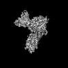 | |||||||||
|---|---|---|---|---|---|---|---|---|---|---|
| Title | Cryo-EM structure of kisspeptin receptor bound to KP-10 | |||||||||
 Map data Map data | ||||||||||
 Sample Sample |
| |||||||||
 Keywords Keywords | Cryo-EM / kisspeptin receptor / GPR54 / KISS1R / G proteins / MEMBRANE PROTEIN | |||||||||
| Function / homology |  Function and homology information Function and homology informationneuropeptide receptor activity / Fatty Acids bound to GPR40 (FFAR1) regulate insulin secretion / Acetylcholine regulates insulin secretion / PLC beta mediated events / phospholipase C-activating dopamine receptor signaling pathway / regulation of platelet activation / phototransduction, visible light / entrainment of circadian clock / G protein-coupled peptide receptor activity / glutamate receptor signaling pathway ...neuropeptide receptor activity / Fatty Acids bound to GPR40 (FFAR1) regulate insulin secretion / Acetylcholine regulates insulin secretion / PLC beta mediated events / phospholipase C-activating dopamine receptor signaling pathway / regulation of platelet activation / phototransduction, visible light / entrainment of circadian clock / G protein-coupled peptide receptor activity / glutamate receptor signaling pathway / regulation of canonical Wnt signaling pathway / action potential / : / photoreceptor outer segment / Adenylate cyclase inhibitory pathway / positive regulation of protein localization to cell cortex / neuropeptide signaling pathway / regulation of cAMP-mediated signaling / D2 dopamine receptor binding / G protein-coupled serotonin receptor binding / regulation of mitotic spindle organization / cellular response to forskolin / adenylate cyclase-inhibiting G protein-coupled receptor signaling pathway / Peptide ligand-binding receptors / GTPase activator activity / Regulation of insulin secretion / G protein-coupled receptor binding / negative regulation of protein kinase activity / G-protein beta/gamma-subunit complex binding / Olfactory Signaling Pathway / Activation of the phototransduction cascade / adenylate cyclase-modulating G protein-coupled receptor signaling pathway / G beta:gamma signalling through PLC beta / Presynaptic function of Kainate receptors / Thromboxane signalling through TP receptor / G-protein activation / G protein-coupled acetylcholine receptor signaling pathway / Activation of G protein gated Potassium channels / Inhibition of voltage gated Ca2+ channels via Gbeta/gamma subunits / adenylate cyclase-activating G protein-coupled receptor signaling pathway / Prostacyclin signalling through prostacyclin receptor / Glucagon signaling in metabolic regulation / G beta:gamma signalling through CDC42 / ADP signalling through P2Y purinoceptor 12 / G beta:gamma signalling through BTK / Synthesis, secretion, and inactivation of Glucagon-like Peptide-1 (GLP-1) / Sensory perception of sweet, bitter, and umami (glutamate) taste / response to peptide hormone / photoreceptor disc membrane / cilium / Adrenaline,noradrenaline inhibits insulin secretion / Glucagon-type ligand receptors / Vasopressin regulates renal water homeostasis via Aquaporins / G alpha (z) signalling events / cellular response to catecholamine stimulus / Glucagon-like Peptide-1 (GLP1) regulates insulin secretion / ADORA2B mediated anti-inflammatory cytokines production / sensory perception of taste / ADP signalling through P2Y purinoceptor 1 / adenylate cyclase-activating dopamine receptor signaling pathway / G beta:gamma signalling through PI3Kgamma / cellular response to prostaglandin E stimulus / Cooperation of PDCL (PhLP1) and TRiC/CCT in G-protein beta folding / GPER1 signaling / GDP binding / G-protein beta-subunit binding / Inactivation, recovery and regulation of the phototransduction cascade / heterotrimeric G-protein complex / G alpha (12/13) signalling events / extracellular vesicle / blood coagulation / signaling receptor complex adaptor activity / Thrombin signalling through proteinase activated receptors (PARs) / GTPase binding / retina development in camera-type eye / phospholipase C-activating G protein-coupled receptor signaling pathway / Ca2+ pathway / cell cortex / midbody / G alpha (i) signalling events / fibroblast proliferation / G alpha (s) signalling events / G alpha (q) signalling events / nuclear membrane / cell population proliferation / Ras protein signal transduction / Extra-nuclear estrogen signaling / protein stabilization / cell cycle / G protein-coupled receptor signaling pathway / lysosomal membrane / cell division / intracellular membrane-bounded organelle / GTPase activity / centrosome / synapse / protein-containing complex binding / nucleolus / GTP binding / Golgi apparatus Similarity search - Function | |||||||||
| Biological species |  Homo Sapiens (human) / Homo sapiens (human) / synthetic construct (others) / Homo Sapiens (human) / Homo sapiens (human) / synthetic construct (others) /  | |||||||||
| Method | single particle reconstruction / cryo EM / Resolution: 3.06 Å | |||||||||
 Authors Authors | Shen S / Liu H / Xu HE | |||||||||
| Funding support |  China, 1 items China, 1 items
| |||||||||
 Citation Citation | Journal: To Be Published Title: Cryo-EM structure of kisspeptin receptor bound to KP-10 Authors: Shen S / Liu H / Xu HE | |||||||||
| History |
|
- Structure visualization
| Supplemental images |
|---|

- Downloads & links
-EMDB archive
| Map data | emd_60141.map.gz | 59.8 MB | EMDB map data format | |
|---|---|---|---|---|
| Header (meta data) | emd-60141-v30.xmlemd-60141.xml | 19 KB 19 KB | Display Display | EMDB header |
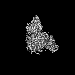
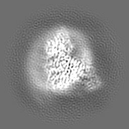
| Images | 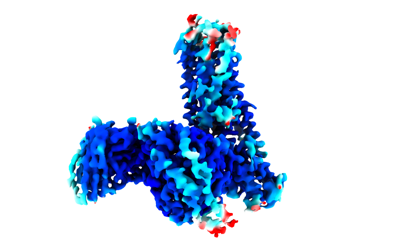 emd_60141.png emd_60141.png | 50.5 KB | ||
| Masks | emd_60141_msk_1.map | 64 MB | Mask map | |
| Filedesc metadata | emd-60141.cif.gz | 6.4 KB | ||
| Others | emd_60141_half_map_1.map.gzemd_60141_half_map_2.map.gz | 59.4 MB 59.4 MB | ||
| Archive directory |  http://ftp.pdbj.org/pub/emdb/structures/EMD-60141ftp://ftp.pdbj.org/pub/emdb/structures/EMD-60141 http://ftp.pdbj.org/pub/emdb/structures/EMD-60141ftp://ftp.pdbj.org/pub/emdb/structures/EMD-60141 | HTTPS FTP |
-Validation report
| Summary document | emd_60141_validation.pdf.gz | 984.3 KB | Display | EMDB validaton report |
|---|---|---|---|---|
| Full document | emd_60141_full_validation.pdf.gz | 983.9 KB | Display | |
| Data in XML | emd_60141_validation.xml.gz | 12.4 KB | Display | |
| Data in CIF | emd_60141_validation.cif.gz | 14.4 KB | Display | |
| Arichive directory | https://ftp.pdbj.org/pub/emdb/validation_reports/EMD-60141ftp://ftp.pdbj.org/pub/emdb/validation_reports/EMD-60141 | HTTPS FTP |
-Related structure data
| Related structure data | 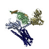 8zjdMC M: atomic model generated by this map C: citing same article ( |
|---|---|
| Similar structure data |
-Links
| EMDB pages | EMDB (EBI/PDBe) / EMDataResource |
|---|---|
| Related items in Molecule of the Month |


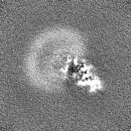
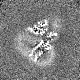
-Map
| File | Download / File: emd_60141.map.gz / Format: CCP4 / Size: 64 MB / Type: IMAGE STORED AS FLOATING POINT NUMBER (4 BYTES) | ||||||||||||||||||||||||||||||||||||
|---|---|---|---|---|---|---|---|---|---|---|---|---|---|---|---|---|---|---|---|---|---|---|---|---|---|---|---|---|---|---|---|---|---|---|---|---|---|
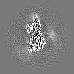
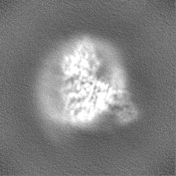
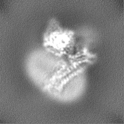
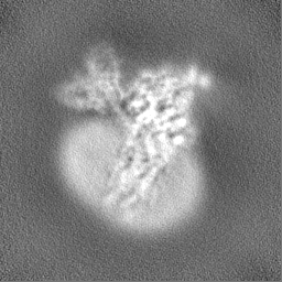
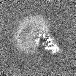
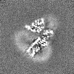
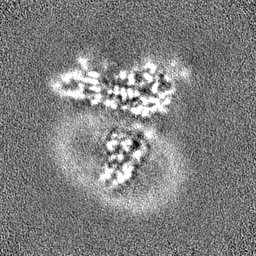
| Projections & slices | Image control
Images are generated by Spider. | ||||||||||||||||||||||||||||||||||||
| Voxel size | X=Y=Z: 0.73 Å | ||||||||||||||||||||||||||||||||||||
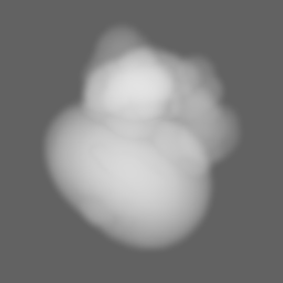
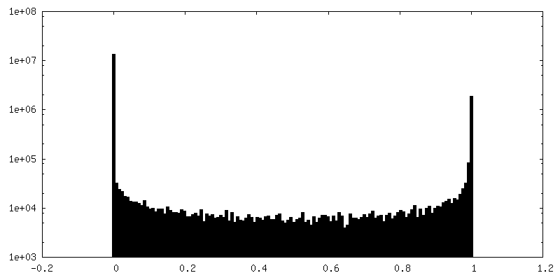
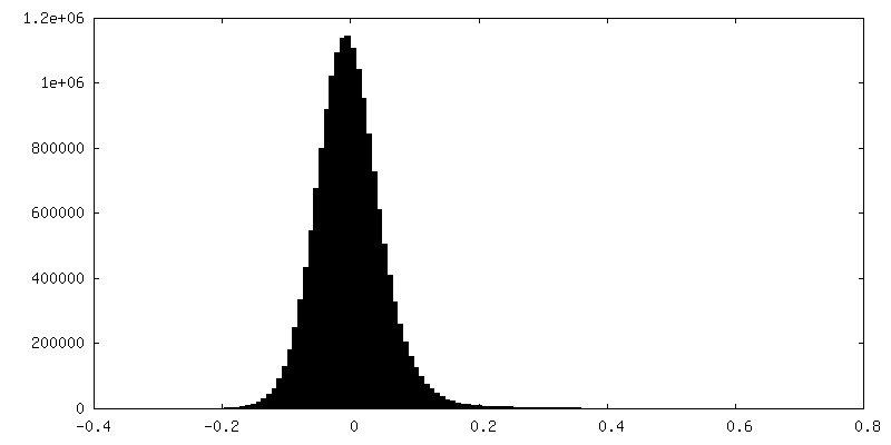
| Density |
| ||||||||||||||||||||||||||||||||||||
| Symmetry | Space group: 1 | ||||||||||||||||||||||||||||||||||||
| Details | EMDB XML:
|
 Z (Sec.)
 Y (Row.)
 X (Col.)


-Supplemental data
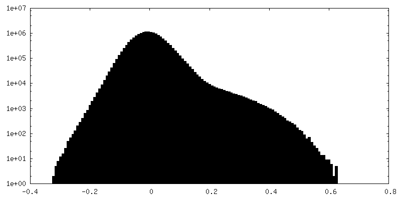
-Mask #1
| File | emd_60141_msk_1.map | ||||||||||||
|---|---|---|---|---|---|---|---|---|---|---|---|---|---|
| Projections & Slices |
| ||||||||||||
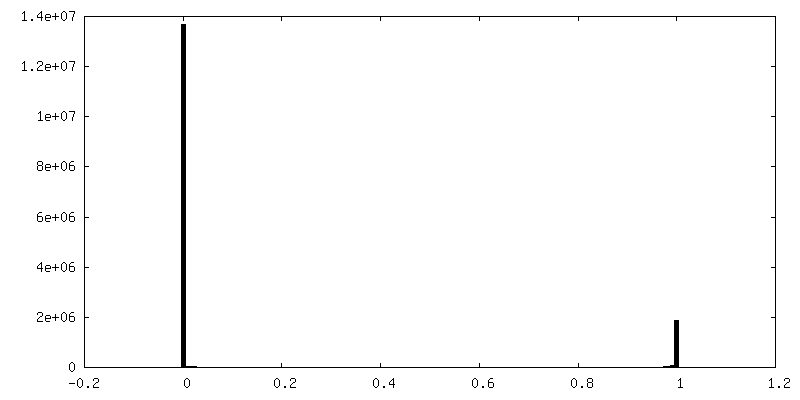
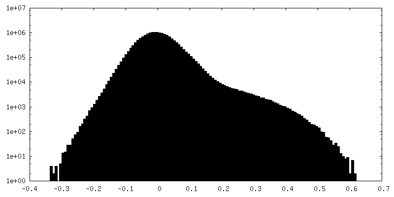
| Density Histograms |


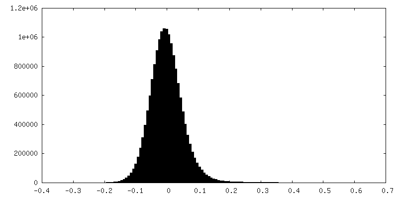
-Half map: #2
| File | emd_60141_half_map_1.map | ||||||||||||
|---|---|---|---|---|---|---|---|---|---|---|---|---|---|
| Projections & Slices |
| ||||||||||||
| Density Histograms |
-Half map: #1
| File | emd_60141_half_map_2.map | ||||||||||||
|---|---|---|---|---|---|---|---|---|---|---|---|---|---|
| Projections & Slices |
| ||||||||||||
| Density Histograms |


- Sample components
-Entire : Cryo-EM structure of kisspeptin receptor in complex with G proteins
| Entire | Name: Cryo-EM structure of kisspeptin receptor in complex with G proteins |
|---|---|
| Components |
|
-Supramolecule #1: Cryo-EM structure of kisspeptin receptor in complex with G proteins
| Supramolecule | Name: Cryo-EM structure of kisspeptin receptor in complex with G proteins type: complex / ID: 1 / Parent: 0 / Macromolecule list: all |
|---|---|
| Source (natural) | Organism: Homo Sapiens (human) |
-Macromolecule #1: KiSS-1 receptor,KiSS-1 receptor,KiSS-1 receptor,KiSS-1 receptor,K...
| Macromolecule | Name: KiSS-1 receptor,KiSS-1 receptor,KiSS-1 receptor,KiSS-1 receptor,KiSS-1 receptor, G-protein coupled receptor 54, GPR54, KISS1R type: protein_or_peptide / ID: 1 / Number of copies: 1 / Enantiomer: LEVO |
|---|---|
| Source (natural) | Organism: Homo sapiens (human) |
| Molecular weight | Theoretical: 55.953941 KDa |
| Recombinant expression | Organism:  Trichoplusia ni (cabbage looper) Trichoplusia ni (cabbage looper) |
| Sequence | String: HTVATSGPNA SWGAPANASG CPGCGANASD GPVPSPRAVD AWLVPLFFAA LMLLGLVGNS LVIYVICRHK PMRTVTNFYI ANLAATDVT FLLCCVPFTA LLYPLPGWVL GDFMCKFVNY IQQVSVQATC WTLTAMSVDR WYVTVFPLRA LHRRTPRLAL A VSLSIWVG ...String: HTVATSGPNA SWGAPANASG CPGCGANASD GPVPSPRAVD AWLVPLFFAA LMLLGLVGNS LVIYVICRHK PMRTVTNFYI ANLAATDVT FLLCCVPFTA LLYPLPGWVL GDFMCKFVNY IQQVSVQATC WTLTAMSVDR WYVTVFPLRA LHRRTPRLAL A VSLSIWVG SAAVSAPVLA LHRLSPGPRA YCSEAFPSRA LERAFALYNL LALYLLPLLA TCACYAAMLR HLGRVAVRPA PA DSALQGQ VLAERAGAVR AKVSRLVAAV VLLFAACWGP IQLFLVLQAL GPAGSWHPRS YAAYALKTWA HCMSYSNSAL NPL LYAFLG SHFRQAFRRV CPCAPRRPRR PRRPGPSDVF TLEDFVGDWE QTAAYNLDQV LEQGGVSSLL QNLAVSVTPI QRIV RSGEN ALKIDIHVII PYEGLSADQM AQIEEVFKVV YPVDDHHFKV ILPYGTLVID GVTPNMLNYF GRPYEGIAVF DGKKI TVTG TLWNGNKIID ERLITPDGSM LFRVTINS UniProtKB: KiSS-1 receptor |
-Macromolecule #2: kisspeptin-10
| Macromolecule | Name: kisspeptin-10 / type: protein_or_peptide / ID: 2 / Number of copies: 1 / Enantiomer: LEVO |
|---|---|
| Source (natural) | Organism: synthetic construct (others) |
| Molecular weight | Theoretical: 1.375511 KDa |
| Sequence | String: YNWNSFGLRF A |
-Macromolecule #3: Guanine nucleotide-binding protein G(I)/G(S)/G(T) subunit beta-1
| Macromolecule | Name: Guanine nucleotide-binding protein G(I)/G(S)/G(T) subunit beta-1 type: protein_or_peptide / ID: 3 / Number of copies: 1 / Enantiomer: LEVO |
|---|---|
| Source (natural) | Organism: Homo sapiens (human) |
| Molecular weight | Theoretical: 38.744371 KDa |
| Recombinant expression | Organism:  Spodoptera frugiperda (fall armyworm) Spodoptera frugiperda (fall armyworm) |
| Sequence | String: MHHHHHHGSL LQSELDQLRQ EAEQLKNQIR DARKACADAT LSQITNNIDP VGRIQMRTRR TLRGHLAKIY AMHWGTDSRL LVSASQDGK LIIWDSYTTN KVHAIPLRSS WVMTCAYAPS GNYVACGGLD NICSIYNLKT REGNVRVSRE LAGHTGYLSC C RFLDDNQI ...String: MHHHHHHGSL LQSELDQLRQ EAEQLKNQIR DARKACADAT LSQITNNIDP VGRIQMRTRR TLRGHLAKIY AMHWGTDSRL LVSASQDGK LIIWDSYTTN KVHAIPLRSS WVMTCAYAPS GNYVACGGLD NICSIYNLKT REGNVRVSRE LAGHTGYLSC C RFLDDNQI VTSSGDTTCA LWDIETGQQT TTFTGHTGDV MSLSLAPDTR LFVSGACDAS AKLWDVREGM CRQTFTGHES DI NAICFFP NGNAFATGSD DATCRLFDLR ADQELMTYSH DNIICGITSV SFSKSGRLLL AGYDDFNCNV WDALKADRAG VLA GHDNRV SCLGVTDDGM AVATGSWDSF LKIWN UniProtKB: Guanine nucleotide-binding protein G(I)/G(S)/G(T) subunit beta-1 |
-Macromolecule #4: scFv16
| Macromolecule | Name: scFv16 / type: protein_or_peptide / ID: 4 / Number of copies: 1 / Enantiomer: LEVO |
|---|---|
| Source (natural) | Organism: |
| Molecular weight | Theoretical: 26.277299 KDa |
| Recombinant expression | Organism: Spodoptera frugiperda (fall armyworm) |
| Sequence | String: VQLVESGGGL VQPGGSRKLS CSASGFAFSS FGMHWVRQAP EKGLEWVAYI SSGSGTIYYA DTVKGRFTIS RDDPKNTLFL QMTSLRSED TAMYYCVRSI YYYGSSPFDF WGQGTTLTVS AGGGGSGGGG SGGGGSADIV MTQATSSVPV TPGESVSISC R SSKSLLHS ...String: VQLVESGGGL VQPGGSRKLS CSASGFAFSS FGMHWVRQAP EKGLEWVAYI SSGSGTIYYA DTVKGRFTIS RDDPKNTLFL QMTSLRSED TAMYYCVRSI YYYGSSPFDF WGQGTTLTVS AGGGGSGGGG SGGGGSADIV MTQATSSVPV TPGESVSISC R SSKSLLHS NGNTYLYWFL QRPGQSPQLL IYRMSNLASG VPDRFSGSGS GTAFTLTISR LEAEDVGVYY CMQHLEYPLT FG AGTKLEL |
-Macromolecule #5: Guanine nucleotide-binding protein G(I)/G(S)/G(O) subunit gamma-2
| Macromolecule | Name: Guanine nucleotide-binding protein G(I)/G(S)/G(O) subunit gamma-2 type: protein_or_peptide / ID: 5 / Number of copies: 1 / Enantiomer: LEVO |
|---|---|
| Source (natural) | Organism: Homo sapiens (human) |
| Molecular weight | Theoretical: 7.861143 KDa |
| Recombinant expression | Organism: Spodoptera frugiperda (fall armyworm) |
| Sequence | String: MASNNTASIA QARKLVEQLK MEANIDRIKV SKAAADLMAY CEAHAKEDPL LTPVPASENP FREKKFFCAI L UniProtKB: Guanine nucleotide-binding protein G(I)/G(S)/G(O) subunit gamma-2 |
-Macromolecule #6: Guanine nucleotide-binding protein G(i) subunit alpha-1,Guanine n...
| Macromolecule | Name: Guanine nucleotide-binding protein G(i) subunit alpha-1,Guanine nucleotide-binding protein G(q) subunit alpha type: protein_or_peptide / ID: 6 Details: fusion protein of Guanine nucleotide-binding protein G(i) subunit alpha-1 and Guanine nucleotide-binding protein G(q) subunit alpha-q. Number of copies: 1 / Enantiomer: LEVO |
|---|---|
| Source (natural) | Organism: Homo sapiens (human) |
| Molecular weight | Theoretical: 41.312922 KDa |
| Recombinant expression | Organism: Spodoptera frugiperda (fall armyworm) |
| Sequence | String: MGCTLSAEDK AAVERSKMID RNLREDGERS RRELKLLLLG TGESGKSTFI KQMRIIHGSG YSDEDKRGFT KLVYQNIFTA MQAMIRAMD TLKIPYKYEH NKAHAQLVRE VDVEKVSAFE NPYVDAIKSL WNDPGIQECY DRRREYQLSD STKYYLNDLD R VADPAYLP ...String: MGCTLSAEDK AAVERSKMID RNLREDGERS RRELKLLLLG TGESGKSTFI KQMRIIHGSG YSDEDKRGFT KLVYQNIFTA MQAMIRAMD TLKIPYKYEH NKAHAQLVRE VDVEKVSAFE NPYVDAIKSL WNDPGIQECY DRRREYQLSD STKYYLNDLD R VADPAYLP TQQDVLRVRV PTTGIIEYPF DLQSVIFRMV DVGAQRSERR KWIHCFENVT SIMFLVALSE YDQVLVESDN EN RMEESKA LFRTIITYPW FQNSSVILFL NKKDLLEEKI MYSHLVDYFP EYDGPQRDAQ AAREFILKMF VDLNPDSDKI IYS HFTCST DTENIRFVFA AVKDTILQLN LKEYNLV UniProtKB: Guanine nucleotide-binding protein G(i) subunit alpha-1, Guanine nucleotide-binding protein G(q) subunit alpha |
-Experimental details
-Structure determination
| Method | cryo EM |
|---|---|
 Processing Processing | single particle reconstruction |
| Aggregation state | particle |
-Sample preparation
| Buffer | pH: 7.4 |
|---|---|
| Vitrification | Cryogen name: ETHANE |
- Electron microscopy
| Microscope | FEI TITAN KRIOS |
|---|---|
| Image recording | Film or detector model: FEI FALCON IV (4k x 4k) / Average electron dose: 50.0 e/Å2 |
| Electron beam | Acceleration voltage: 300 kV / Electron source:  FIELD EMISSION GUN FIELD EMISSION GUN |
| Electron optics | Illumination mode: FLOOD BEAM / Imaging mode: DARK FIELD / Nominal defocus max: 2.0 µm / Nominal defocus min: 1.0 µm |
| Experimental equipment |  Model: Titan Krios / Image courtesy: FEI Company |
-Image processing
| Startup model | Type of model: NONE |
|---|---|
| Final reconstruction | Resolution.type: BY AUTHOR / Resolution: 3.06 Å / Resolution method: FSC 0.143 CUT-OFF / Number images used: 199591 |
| Initial angle assignment | Type: MAXIMUM LIKELIHOOD |
| Final angle assignment | Type: MAXIMUM LIKELIHOOD |
