[English] 日本語
 Yorodumi
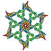
- EMDB-51414: Surface-layer (S-layer) PS2 protein from Corynebacterium glutamicum -
+ Open data
- Basic information
| Entry |  | |||||||||
|---|---|---|---|---|---|---|---|---|---|---|
| Title | Surface-layer (S-layer) PS2 protein from Corynebacterium glutamicum | |||||||||
 Map data Map data | ||||||||||
 Sample Sample |
| |||||||||
 Keywords Keywords | S-layer / Corynebacterium / Surface / STRUCTURAL PROTEIN | |||||||||
| Function / homology | membrane / PS2 Function and homology information Function and homology information | |||||||||
| Biological species |  Corynebacterium glutamicum (bacteria) Corynebacterium glutamicum (bacteria) | |||||||||
| Method | single particle reconstruction / cryo EM / Resolution: 2.5 Å | |||||||||
 Authors Authors | Sogues A / Remaut H / Sleutel M | |||||||||
| Funding support | European Union, 2 items
| |||||||||
 Citation Citation |  Journal: bioRxiv / Year: 2024 Journal: bioRxiv / Year: 2024Title: Cryo-EM structure and polar assembly of the PS2 S-layer of . Authors: Adrià Sogues / Mike Sleutel / Julienne Petit / Daniela Megrian / Nicolas Bayan / Anne Marie Wehenkel / Han Remaut /   Abstract: The polar-growing Corynebacteriales have a complex cell envelope architecture characterized by the presence of a specialized outer membrane composed of mycolic acids. In some Corynebacteriales, this ...The polar-growing Corynebacteriales have a complex cell envelope architecture characterized by the presence of a specialized outer membrane composed of mycolic acids. In some Corynebacteriales, this mycomembrane is further supported by a proteinaceous surface layer or 'S-layer', whose function, structure and mode of assembly remain largely enigmatic. Here, we isolated PS2 S-layers from the industrially important and determined its atomic structure by 3D cryoEM reconstruction. PS2 monomers consist of a six-helix bundle 'core', a three-helix bundle 'arm', and a C-terminal transmembrane (TM) helix. The PS2 core oligomerizes into hexameric units anchored in the mycomembrane by a channel-like coiled-coil of the TM helices. The PS2 arms mediate trimeric lattice contacts, crystallizing the hexameric units into an intricate semipermeable lattice. Using pulse-chase live cell imaging, we show that the PS2 lattice is incorporated at the poles, coincident with the actinobacterial elongasome. Finally, phylogenetic analysis shows a paraphyletic distribution and dispersed chromosomal location of PS2 in Corynebacteriales as a result of multiple recombination events and losses. These findings expand our understanding of S-layer biology and enable applications of membrane-supported self-assembling bioengineered materials. | |||||||||
| History |
|
- Structure visualization
| Supplemental images |
|---|
- Downloads & links
-EMDB archive
| Map data |  emd_51414.map.gz emd_51414.map.gz | 408.1 MB |  EMDB map data format EMDB map data format | |
|---|---|---|---|---|
| Header (meta data) |  emd-51414-v30.xml emd-51414-v30.xml emd-51414.xml emd-51414.xml | 13.5 KB 13.5 KB | Display Display |  EMDB header EMDB header |
| FSC (resolution estimation) |  emd_51414_fsc.xml emd_51414_fsc.xml | 19.8 KB | Display |  FSC data file FSC data file |
| Images |  emd_51414.png emd_51414.png | 155.6 KB | ||
| Masks |  emd_51414_msk_1.map emd_51414_msk_1.map | 824 MB |  Mask map Mask map | |
| Filedesc metadata |  emd-51414.cif.gz emd-51414.cif.gz | 5.2 KB | ||
| Others |  emd_51414_half_map_1.map.gz emd_51414_half_map_1.map.gz emd_51414_half_map_2.map.gz emd_51414_half_map_2.map.gz | 765.3 MB 765.2 MB | ||
| Archive directory |  http://ftp.pdbj.org/pub/emdb/structures/EMD-51414 http://ftp.pdbj.org/pub/emdb/structures/EMD-51414 ftp://ftp.pdbj.org/pub/emdb/structures/EMD-51414 ftp://ftp.pdbj.org/pub/emdb/structures/EMD-51414 | HTTPS FTP |
-Validation report
| Summary document |  emd_51414_validation.pdf.gz emd_51414_validation.pdf.gz | 1.1 MB | Display |  EMDB validaton report EMDB validaton report |
|---|---|---|---|---|
| Full document |  emd_51414_full_validation.pdf.gz emd_51414_full_validation.pdf.gz | 1.1 MB | Display | |
| Data in XML |  emd_51414_validation.xml.gz emd_51414_validation.xml.gz | 29.5 KB | Display | |
| Data in CIF |  emd_51414_validation.cif.gz emd_51414_validation.cif.gz | 39 KB | Display | |
| Arichive directory |  https://ftp.pdbj.org/pub/emdb/validation_reports/EMD-51414 https://ftp.pdbj.org/pub/emdb/validation_reports/EMD-51414 ftp://ftp.pdbj.org/pub/emdb/validation_reports/EMD-51414 ftp://ftp.pdbj.org/pub/emdb/validation_reports/EMD-51414 | HTTPS FTP |
-Related structure data
| Related structure data |  9gk2MC M: atomic model generated by this map C: citing same article ( |
|---|---|
| Similar structure data | Similarity search - Function & homology  F&H Search F&H Search |
- Links
Links
| EMDB pages |  EMDB (EBI/PDBe) / EMDB (EBI/PDBe) /  EMDataResource EMDataResource |
|---|
- Map
Map
| File |  Download / File: emd_51414.map.gz / Format: CCP4 / Size: 824 MB / Type: IMAGE STORED AS FLOATING POINT NUMBER (4 BYTES) Download / File: emd_51414.map.gz / Format: CCP4 / Size: 824 MB / Type: IMAGE STORED AS FLOATING POINT NUMBER (4 BYTES) | ||||||||||||||||||||||||||||||||||||
|---|---|---|---|---|---|---|---|---|---|---|---|---|---|---|---|---|---|---|---|---|---|---|---|---|---|---|---|---|---|---|---|---|---|---|---|---|---|
| Projections & slices | Image control
Images are generated by Spider. | ||||||||||||||||||||||||||||||||||||
| Voxel size | X=Y=Z: 0.71 Å | ||||||||||||||||||||||||||||||||||||
| Density |
| ||||||||||||||||||||||||||||||||||||
| Symmetry | Space group: 1 | ||||||||||||||||||||||||||||||||||||
| Details | EMDB XML:
|
-Supplemental data
-Mask #1
| File |  emd_51414_msk_1.map emd_51414_msk_1.map | ||||||||||||
|---|---|---|---|---|---|---|---|---|---|---|---|---|---|
| Projections & Slices |
| ||||||||||||
| Density Histograms |
-Half map: #1
| File | emd_51414_half_map_1.map | ||||||||||||
|---|---|---|---|---|---|---|---|---|---|---|---|---|---|
| Projections & Slices |
| ||||||||||||
| Density Histograms |
-Half map: #2
| File | emd_51414_half_map_2.map | ||||||||||||
|---|---|---|---|---|---|---|---|---|---|---|---|---|---|
| Projections & Slices |
| ||||||||||||
| Density Histograms |
- Sample components
-Entire : Ex vivo PS2 S-layer
| Entire | Name: Ex vivo PS2 S-layer |
|---|---|
| Components |
|
-Supramolecule #1: Ex vivo PS2 S-layer
| Supramolecule | Name: Ex vivo PS2 S-layer / type: organelle_or_cellular_component / ID: 1 / Parent: 0 / Macromolecule list: all |
|---|---|
| Source (natural) | Organism:  Corynebacterium glutamicum (bacteria) Corynebacterium glutamicum (bacteria) |
-Macromolecule #1: PS2
| Macromolecule | Name: PS2 / type: protein_or_peptide / ID: 1 / Number of copies: 18 / Enantiomer: LEVO |
|---|---|
| Source (natural) | Organism:  Corynebacterium glutamicum (bacteria) Corynebacterium glutamicum (bacteria) |
| Molecular weight | Theoretical: 47.666859 KDa |
| Recombinant expression | Organism:  Corynebacterium glutamicum (bacteria) Corynebacterium glutamicum (bacteria) |
| Sequence | String: ETNPTFNITN GFNDADGSTI QPVGPVNHTE ETLRDLTDST GAYLEEFQNG TVEEIVEAYL QVQASADGFD PSEQAAYEAF EAARVRASQ ELAASAETIT KTRESVAYAL KVDQEATAAF EAYRNALRDA AISINPDGSI NPDTSINLLI DAANAANRTD R AEIEDYAH ...String: ETNPTFNITN GFNDADGSTI QPVGPVNHTE ETLRDLTDST GAYLEEFQNG TVEEIVEAYL QVQASADGFD PSEQAAYEAF EAARVRASQ ELAASAETIT KTRESVAYAL KVDQEATAAF EAYRNALRDA AISINPDGSI NPDTSINLLI DAANAANRTD R AEIEDYAH LYTQTDIALE TPQLAYAFQD LKALQAEVDA DFEWLGEFGI DQEDGNYVQR YHLPAVEALK AEVDARVAAI EP LRADSIA KNLEAQKSDV LVRQLFLERA TAQRDTLRVV EAIFSTSARY VELYENVENV NVENKTLRQH YSALIPNLFI AAV ANISEL NAADAEAAAY YLHWDTDLAT NDEDEAYYKA KLDFAIETYA KILFNGEVWQ EPLAYVQNLD AGARQEAADR EAAR AADEA YRAEQLRIAQ EAADAQKAIA EALAKEA UniProtKB: PS2 |
-Experimental details
-Structure determination
| Method | cryo EM |
|---|---|
 Processing Processing | single particle reconstruction |
| Aggregation state | 2D array |
- Sample preparation
| Buffer | pH: 7.4 |
|---|---|
| Vitrification | Cryogen name: ETHANE |
- Electron microscopy
| Microscope | JEOL CRYO ARM 300 |
|---|---|
| Image recording | Film or detector model: GATAN K3 (6k x 4k) / Average electron dose: 60.3 e/Å2 |
| Electron beam | Acceleration voltage: 300 kV / Electron source:  FIELD EMISSION GUN FIELD EMISSION GUN |
| Electron optics | Illumination mode: FLOOD BEAM / Imaging mode: BRIGHT FIELD / Nominal defocus max: 1.6 µm / Nominal defocus min: 1.4000000000000001 µm |
 Movie
Movie Controller


 Z (Sec.)
 Y (Row.)
 X (Col.)


